. 2023 Apr 21;14(1):2285.
doi: 10.1038/s41467-023-37674-8.

Immune cell dynamics deconvoluted by single-cell RNA sequencing in normothermic machine perfusion of the liver

Affiliations

Immune cell dynamics deconvoluted by single-cell RNA sequencing in normothermic machine perfusion of the liver

T Hautz et al. Nat Commun. .

Abstract

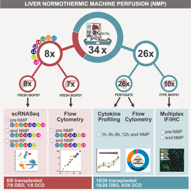
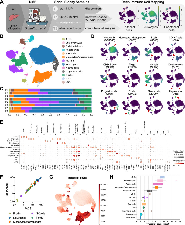
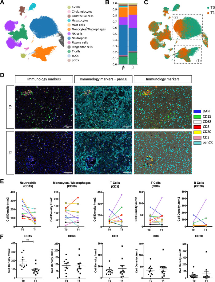
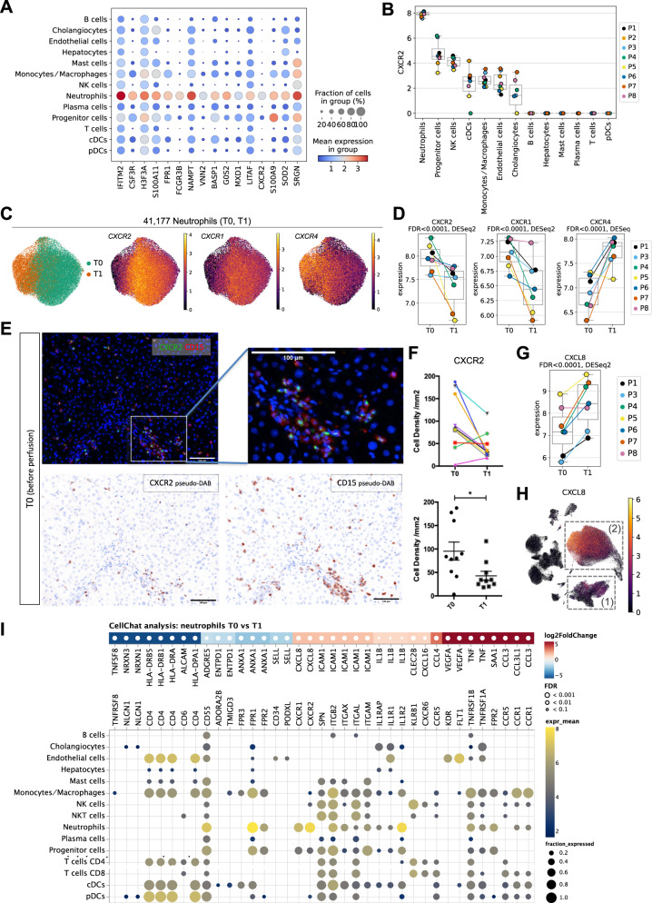
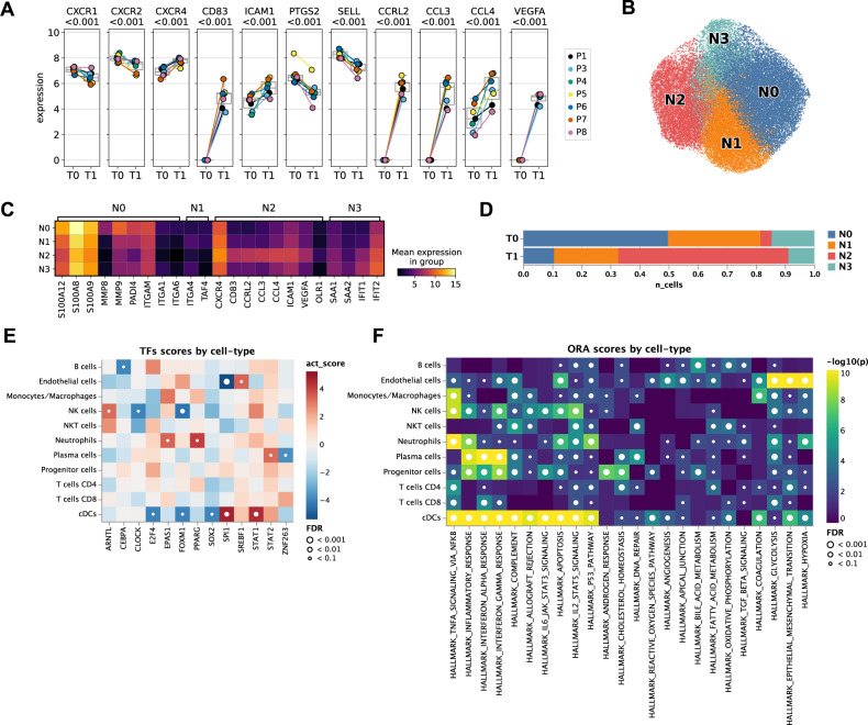
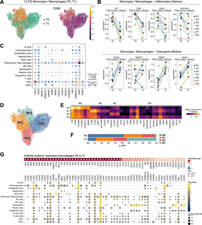
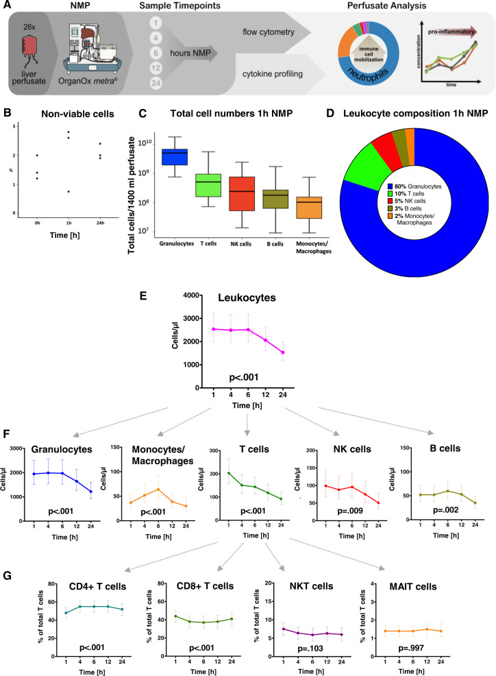
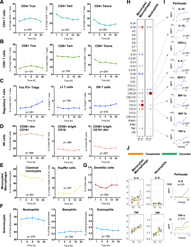
Normothermic machine perfusion (NMP) has emerged as an innovative organ preservation technique. Developing an understanding for the donor organ immune cell composition and its dynamic changes during NMP is essential. We aimed for a comprehensive characterization of immune cell (sub)populations, cell trafficking and cytokine release during liver NMP. Single-cell transcriptome profiling of human donor livers prior to, during NMP and after transplantation shows an abundance of CXC chemokine receptor 1+/2+ (CXCR1+/CXCR2+) neutrophils, which significantly decreased during NMP. This is paralleled by a large efflux of passenger leukocytes with neutrophil predominance in the perfusate. During NMP, neutrophils shift from a pro-inflammatory state towards an aged/chronically activated/exhausted phenotype, while anti-inflammatory/tolerogenic monocytes/macrophages are increased. We herein describe the dynamics of the immune cell repertoire, phenotypic immune cell shifts and a dominance of neutrophils during liver NMP, which potentially contribute to the inflammatory response. Our findings may serve as resource to initiate future immune-interventional studies.


Conflict of interest statement

The authors declare no competing interests.

Figures

Fig. 1
Fig. 1. Overview of study.
While a total of 34 donor livers were included in this study and subjected to NMP, eight out of 34 donor livers were randomly selected for scRNASeq, and 26 out of 34 for perfusate sampling. In addition, tissue samples were collected in all 34 livers to assess and validate findings on protein level. Information is provided on the sampling time points, analysis, donor as well as transplantation status of the organs. FFPE fresh frozen paraffin-embedded, NMP normothermic machine perfusion, RP reperfusion, IF immunofluorescence, IHC immunohistochemistry, DBD donor after brain death, DCD donor after circulatory death.
Fig. 2
Fig. 2. scRNASeq profiling of liver allografts.
A Overview of the scRNASeq workflow. The proportions of epithelial cells (KRT18), leukocytes (PTPRC/CD45), and endothelial cells (SPARC) in the obtained scRNASeq dataset are indicated. B Uniform manifold approximation and projection (UMAP) plot of 118,448 single cells, color-coded by cell type. C Relative cell-type composition in liver tissues from eight individual patients. D UMAP plots, color-coded for the expression of indicated cell-type specific marker genes (red arrowheads). E Gene-expression levels of cell-type specific markers. F The proportion of leukocyte populations in liver tissue as determined by scRNASeq vs flow cytometry. G UMAP plot colored by the number of transcripts per cell. The color scale is clipped at 20,000. H Boxplot of the transcript count per cell-type. The values denote the average per patient (n = 8). The central line denotes the median. Boxes represent the interquartile range (IQR) of the data, whiskers extend to the most extreme data points within 1.5 times the IQR.
Fig. 3
Fig. 3. Impact of NMP on immune cells within the liver.
A UMAP plot of 90,404 single cells (only T0 and T1 samples), color-coded by cell type. B Relative cell type composition in liver tissues pre (T0) and at the end of (T1) NMP. C UMAP-plot of 90,404 single-cells, color-coded by time-point [pre (T0) and at the end of (T1) NMP]. The monocyte/macrophage (1) and neutrophil (2) clusters are highlighted. D Multiplex Immunofluorescence images of immunology markers presented alone and together with pan-Cytokeratin and the phenotyping map pre (T0) and at the end of (T1) NMP. The phenotyping map was generated in InForm, each color dot represents a phenotype of the cell, black dots: other cells of unknown phenotype. Images are displayed at ×20 magnification (scale bar: 100 µm). E, F Cell densities (number of cells/mm2) for individual immunology markers in 10 patients. The upper panel E presents the values for each individual liver pre (T0) and at the end of (T1) NMP. The lower panel F presents a column statistical analysis for each biomarker (n = 10, paired t-test, two-tailed, mean ± SEM, **p = 0.0097).
Fig. 4
Fig. 4. Impact of NMP on neutrophils.
A Gene expression levels of neutrophil-specific genes in individual cell types. B CXCR2 gene expression levels in individual cell types. Each dot represents the mean of a patient (n = 8). C UMAP plots of 41,177 neutrophils, color-coded by timepoint and the CXCR2, CXCR1, and CXCR4 gene expression levels. D CXCR2, CXCR1, and CXCR4 gene expression levels in neutrophils pre (T0) and at the end of (T1) NMP. Each dot represents the mean of a patient (n = 7). False-discovery rates (FDR) have been determined using pseudo-bulk analysis with DESeq2. E Immunofluorescent staining of CXCR2 and CD15 pre (T0) NMP. Images are displayed at ×20 magnification (scale bar: 100 µm, n = 10, for each sample 4 representative images were taken). F Cell density (number of cells/mm2) of CXCR2+ cells pre (T0) and at the end of (T1) NMP. The upper graph presents the values for each individual liver, the lower graph is a column statistical analysis (n = 10, paired t-test, two-tailed, mean ± SEM, *p = 0.02). G CXCL8 gene expression level in neutrophils pre (T0) and at the end of (T1) NMP. Each dot represents the mean of a patient (n = 7). False-discovery rates (FDR) have been determined using pseudo-bulk analysis with DESeq2. H UMAP plot of 90,404 single cells (only T0 and T1 samples), colored by CXCL8 gene expression. The monocyts/macrophage (1) and neutrophil (2) clusters are highlighted. I Differential signaling from neutrophils to other cell types. Upper panel: Differentially expressed ligands of neutrophils in T0 vs. T1 (two-sided DESeq2 Wald-test on pseudo-bulk, p-values were adjusted to false-discovery rate (FDR)). Red colors indicate upregulation in T1 compared to T0. Lower panel: Respective receptors and the expression by cell type. Dot sizes and colors refer to the fraction of cells expressing the receptor and gene expression, respectively, averaged over all patients. Dots are only shown for receptors that are expressed in at least 10% of the respective cell types. In all boxplots, the central line denotes the median. Boxes represent the interquartile range (IQR) of the data, whiskers extend to the most extreme data points within 1.5 times the IQR.
Fig. 5
Fig. 5. NMP shifts neutrophils towards an aged/chronically activated/exhausted phenotype.
A Expression of selected marker genes in neutrophils pre (T0) and at the end of (T1) NMP in individual patients (n = 7). The central line denotes the median. Boxes represent the interquartile range (IQR) of the data, whiskers extend to the most extreme data points within 1.5 times the IQR. Two-sided DESeq2 Wald-test on pseudo-bulk, p-values are adjusted to false-discovery rate (FDR) using independent hypothesis weighting (IHW). B UMAP of neutrophils colored by subclusters (N0, N1, N2, N3). C Expression of selected marker genes in neutrophil subclusters. D Relative composition of neutrophil subclusters pre (T0) and at the end of (T1) NMP. E Differential transcription factor activity (TF) activity per cell-type in T0 vs. T1 computed using DoRothEA. Red indicates upregulation of a TF-regulon in T1 compared to T0. p-values were determined using a two-sided t-test and adjusted for false-discovery rate (FDR). The t-statistics were computed using a multivariate linear model as implemented in decoupler-py. F Gene set over-representation analysis (ORA) of selected “Hallmark” gene sets per cell type in T0 vs. T1. A small p-value indicates that among genes in the gene set, more genes are differentially expressed than expected by chance (one-tailed Fisher’s exact test).
Fig. 6
Fig. 6. Impact of NMP on monocytes/macrophages.
A UMAP-plots of 13,720 monocytes/macrophages, color-coded by time-point [pre (T0) and at the end of (T1) NMP], and of the relative CD68 gene expression level. B Gene expression levels of inflammatory markers (LYZ, FCN1, VCAN, HLA-DRA) and tolerogenic markers (CD163, MARCO, HMOX1, and VSIG4) in monocytes/macrophages pre (T0) and at the end of NMP (T1). Each dot represents the mean of a patient (n = 7). False-discovery rates (FDR) have been determined using pseudo-bulk analysis with DESeq2. The central line denotes the median. Boxes represent the interquartile range (IQR) of the data, whiskers extend to the most extreme data points within 1.5 times the IQR. C Gene expression levels of monocyte/macrophage-specific genes. D UMAP of monocytes/macrophages colored by subclusters (M0, M1, M2, M3). E Expression of selected marker genes in monocyte/macrophage subclusters. F Relative composition of monocyte/macrophage subclusters pre (T0) and at the end of (T1) NMP. G Differential signaling from monocytes/macrophages to other cell-types. Upper panel: Differentially expressed ligands of monocytes/macrophages in T0 vs. T1 (two-sided DESeq2 Wald-test on pseudo-bulk, p-values were adjusted to false-discovery rate (FDR)). Red colors indicate upregulation in T1 compared to T0. Lower panel: Respective receptors and the expression by cell type. Dot sizes and colors refer to the fraction of cells expressing the receptor and gene expression, respectively, averaged over all patients. Dots are only shown for receptors that are expressed in at least 10% of the respective cell types. Only upregulated ligands are shown. The list of downregulated interactions is available in the Source Data file.
Fig. 7
Fig. 7. Total release of passenger leukocytes into the perfusate (n = 26) at various time points during NMP.
A NMP perfusate analysis workflow and applied methods. B Cell viability testing prior to flow cytometry (n = 3 pre, at 1 h NMP and at the end of NMP) showed only a very small percentage of non-viable cells in the perfusate. C Absolute CD45+ leukocyte amounts for main immune cell subtypes in total circulating perfusate (mean ± SEM) and D composition at 1 h NMP. E, F Dynamic change of total CD45+ leukocytes and main subtypes during NMP. G Dynamic change of CD3+ T cell subtypes (proportions) over perfusion time. Graphs show the marginal effects. The values are estimated using linear regression analysis. The p-values refer to the change over time. The least-squares means computed using a linear model are shown together with the 95% CI. N = 26 biologically independent samples. Source data are provided as a Source Data file. NK cells natural killer cells, NKT cells natural killer T cells, MAIT cells mucosa-associated invariant T cells.
Fig. 8
Fig. 8. Phenotyping of perfusate immune cells and inflammatory profile during NMP.
Perfusates collected at various time points during NMP (n = 26 livers subjected to NMP) were assessed for their dynamic changes of the proportions of A CD4+ T cells, B CD8+ T cells, C subtypes of CD3+ T cells with regulatory properties, D CD56+ NK cells, E CD45+CD14+ monocytes/macrophages, F CD45+HLA-DRlow granulocytes, and G dendritic cells. Graphs show the marginal effects. The values are estimated using linear regression analysis. The p-values refer to the change over time. The Least-Squares Means computed using a linear model is shown together with the 95% CI. N = 26 biologically independent samples. H Gene expression levels of indicated pro-inflammatory factors (interleukins/chemokines) in monocytes/macrophages and neutrophils pre (T0) and at the end of NMP (T1) as assessed by scRNASeq in eight donor livers. I Spectrum of assessed pro-inflammatory interleukins/chemokines produced by monocytes/macrophages and neutrophils and elevated on protein level in perfusate samples collected over perfusion time of 26 donor livers. (J) Gene expression levels of IL-6 and TNF in monocytes/macrophages and neutrophils pre (T0) and at the end of NMP (T1) as assessed by scRNASeq in transplanted (n = 6) and discarded (n = 2) livers. The central line denotes the median. Boxes represent the interquartile range (IQR) of the data, whiskers extend to the most extreme data points within 1.5 times the IQR. In addition, the perfusate of 26 donor livers subjected to NMP was measured for IL-6 and TNF levels at 1, 4, 6, 12 and 24 h of NMP and differences between transplanted (n = 18) and discarded (n = 8) livers were calculated. Graphs show the marginal effects. The values are estimated using linear regression analysis. The p-values refer to the change over time. The Least Squares Means computed using a linear model are shown together with the 95% CI. N = 26 biologically independent samples. Source data are provided as a Source Data file. Tcm: central memory T cells, Tem: effector memory T cells, Temra: effector memory cells re-expressing CD45RA T cells, DN T cells: double-negative T cells (CD4 and CD8-), NK cells Natural killer cells, DCs dendritic cells.

References

    1. Cardini B, et al. Live confocal imaging as a novel tool to assess liver quality: insights from a murine model. Transplantation. 2020;104:2528–2537. doi: 10.1097/TP.0000000000003405. - DOI - PubMed
    1. Cardini B, et al. Clinical implementation of prolonged liver preservation and monitoring through normothermic machine perfusion in liver transplantation. Transplantation. 2020;104:1917–1928. doi: 10.1097/TP.0000000000003296. - DOI - PubMed
    1. Fodor M, et al. The need to update endpoints and outcome analysis in the rapidly changing field of liver transplantation. Transplantation. 2021;106:938–949. doi: 10.1097/TP.0000000000003973. - DOI - PubMed
    1. Nasralla D, et al. A randomized trial of normothermic preservation in liver transplantation. Nature. 2018;557:50–56. doi: 10.1038/s41586-018-0047-9. - DOI - PubMed
    1. Ceresa CDL, Nasralla D, Coussios CC, Friend PJ. The case for normothermic machine perfusion in liver transplantation. Liver Transpl. 2018;24:269–275. doi: 10.1002/lt.25000. - DOI - PubMed

Publication types