. 2023 Apr 21;14(4):284.
doi: 10.1038/s41419-023-05780-6.

TRAP1 S-nitrosylation as a model of population-shift mechanism to study the effects of nitric oxide on redox-sensitive oncoproteins

Affiliations

TRAP1 S-nitrosylation as a model of population-shift mechanism to study the effects of nitric oxide on redox-sensitive oncoproteins

Elena Papaleo et al. Cell Death Dis. .

Abstract

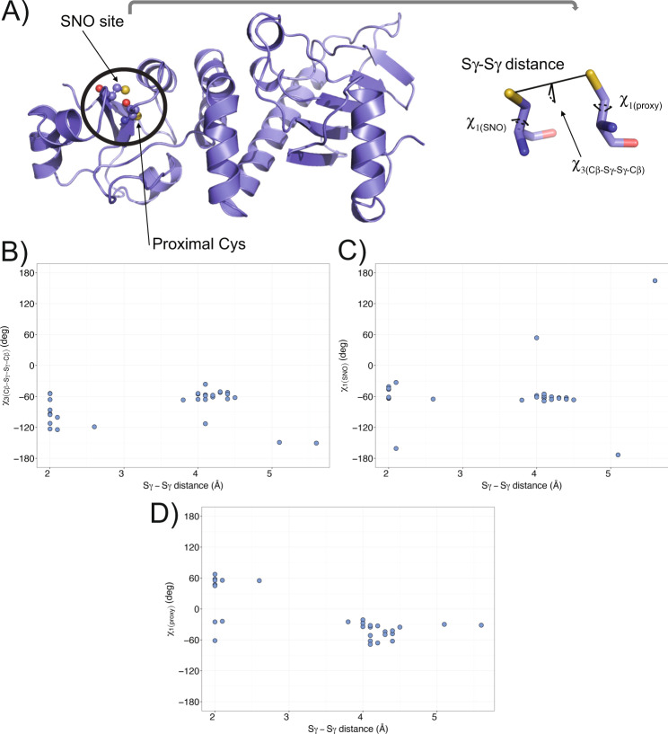
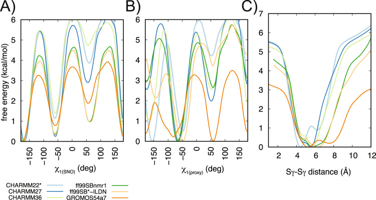
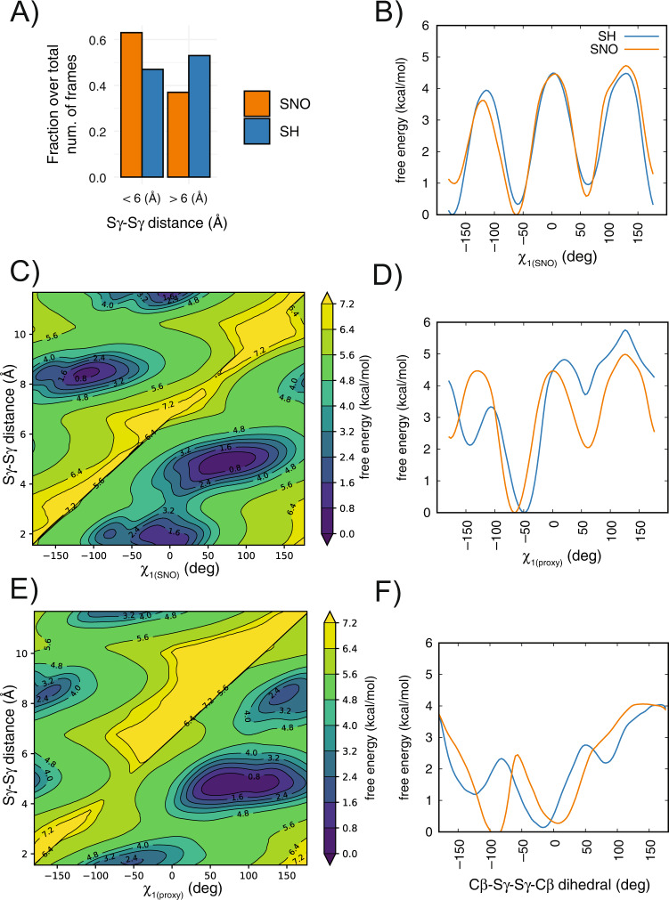
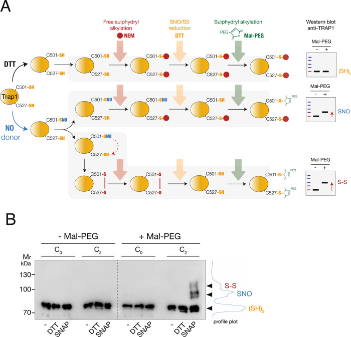
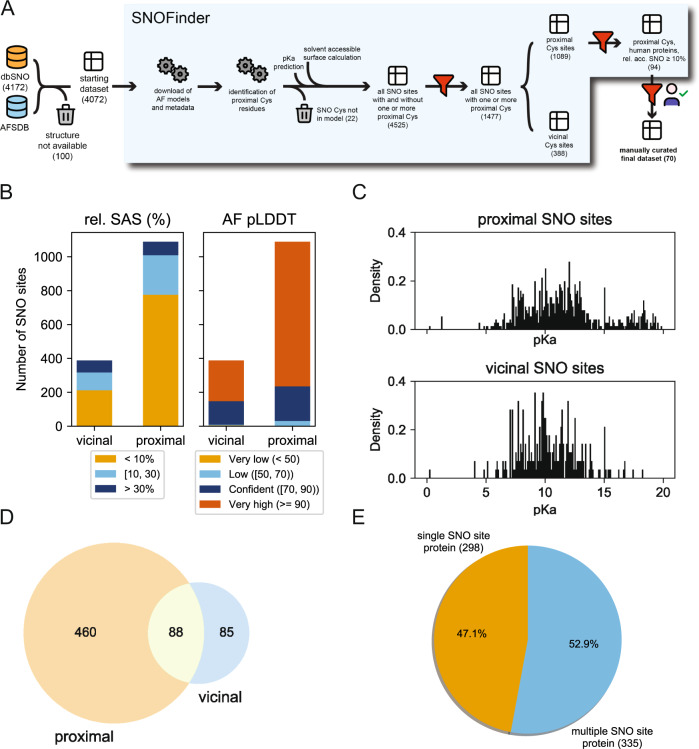
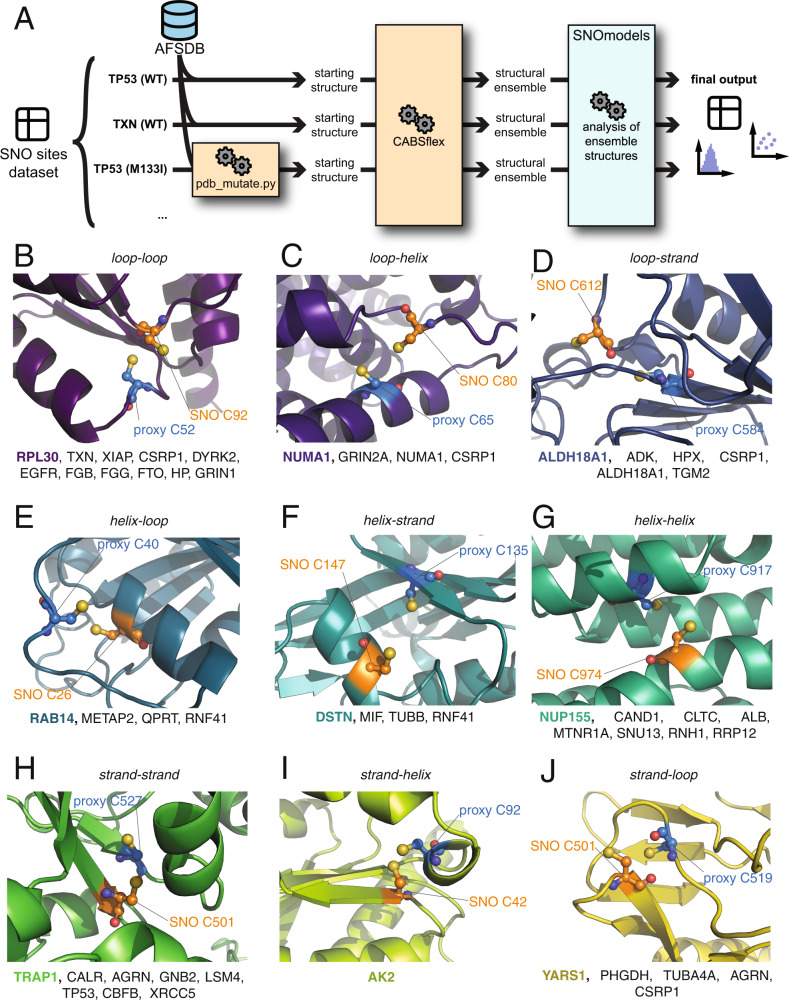
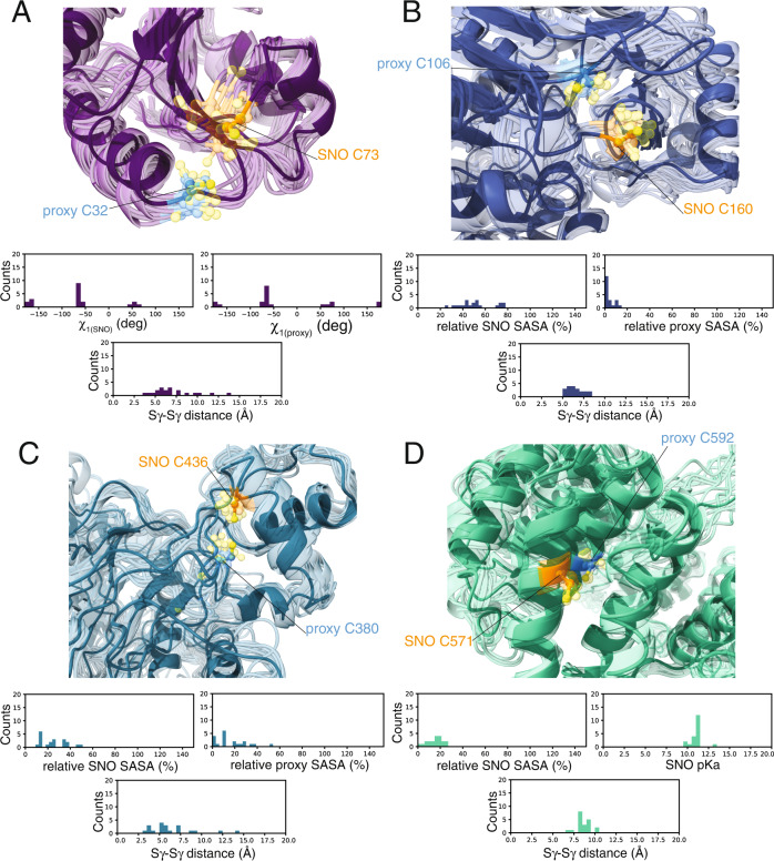
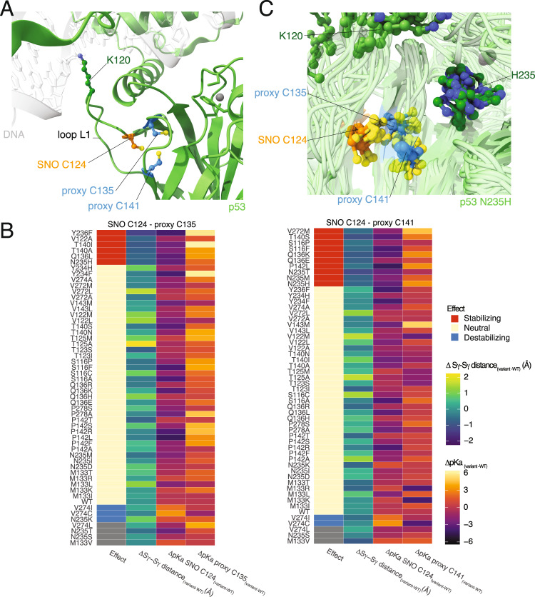
S-nitrosylation is a post-translational modification in which nitric oxide (NO) binds to the thiol group of cysteine, generating an S-nitrosothiol (SNO) adduct. S-nitrosylation has different physiological roles, and its alteration has also been linked to a growing list of pathologies, including cancer. SNO can affect the function and stability of different proteins, such as the mitochondrial chaperone TRAP1. Interestingly, the SNO site (C501) of TRAP1 is in the proximity of another cysteine (C527). This feature suggests that the S-nitrosylated C501 could engage in a disulfide bridge with C527 in TRAP1, resembling the well-known ability of S-nitrosylated cysteines to resolve in disulfide bridge with vicinal cysteines. We used enhanced sampling simulations and in-vitro biochemical assays to address the structural mechanisms induced by TRAP1 S-nitrosylation. We showed that the SNO site induces conformational changes in the proximal cysteine and favors conformations suitable for disulfide bridge formation. We explored 4172 known S-nitrosylated proteins using high-throughput structural analyses. Furthermore, we used a coarse-grained model for 44 protein targets to account for protein flexibility. This resulted in the identification of up to 1248 proximal cysteines, which could sense the redox state of the SNO site, opening new perspectives on the biological effects of redox switches. In addition, we devised two bioinformatic workflows ( https://github.com/ELELAB/SNO_investigation_pipelines ) to identify proximal or vicinal cysteines for a SNO site with accompanying structural annotations. Finally, we analyzed mutations in tumor suppressors or oncogenes in connection with the conformational switch induced by S-nitrosylation. We classified the variants as neutral, stabilizing, or destabilizing for the propensity to be S-nitrosylated and undergo the population-shift mechanism. The methods applied here provide a comprehensive toolkit for future high-throughput studies of new protein candidates, variant classification, and a rich data source for the research community in the NO field.


Conflict of interest statement

The authors declare no competing interests.

Figures

Fig. 1
Fig. 1. Conformational heterogeneity of the S-nitrosylated cysteine and its proximal cysteine in the mitochondrial chaperone TRAP1.
A In the left panel, the cartoon shows the X-ray structure of the middle domain of Danio rerio TRAP1 (PDB ID entry 4IPE, residues 311–567). The spheres indicate the atoms of the S-nitrosylated (SNO) site (C516) and the proximal cysteine (C542). In the right panel, the stick representation shows a conformation of the SNO site and the proximal cysteine. We evaluated the conformational state and orientation of each cysteine, measuring i) the χ1 dihedral angle of the SNO cysteine (χ1(SNO)), ii) the χ1 dihedral angle of the proximal cysteine (χ1(proxy)), iii) the distance between the two sulfur atoms of the two cysteines (Sγ-Sγ distance), and iv) the Cβ-Sγ-Sγ-Cβ dihedral angle. BD We retrieved from the Protein Data Bank (PDB) the structures available for Homo Sapiens and Danio rerio TRAP1. We evaluated the conformational state of the S-nitrosylated cysteine (C501 and C516, respectively) and its proximal cysteine (C527 and C542, respectively). The scatter plots show the calculated values for each structure of the Sγ-Sγ distance against (B) Cβ-Sγ-Sγ-Cβ, (C) χ1(SNO), and (D) χ1(proxy) dihedral angles. We observed heterogeneity in the conformation and orientation of the two cysteines, showing Sγ-Sγ distances in the range of 2–5.6 Å and diversity in Cβ-Sγ-Sγ-Cβ (in the range between −150.5 and −36.2°), χ1(SNO) (in the range between −173.3 and 164.5°), and χ1(proxy) (in the range between −68.7 and 55.0°) dihedral angles.
Fig. 2
Fig. 2. Different MD force fields agree on the conformational preferences of the reduced form of the two cysteines of TRAP1311–567.
The panels show the mono-dimensional free energy profiles associated with the collective variables (A) χ1(SNO), (B) χ1(proxy) and (C) Sγ-Sγ distance for the metadynamics simulations. We performed metadynamics using CHARMM22* (light blue), CHARMM27 (dark blue), CHARMM36 (light green), ff99SBnmr1 (dark green), ff99SB*-ILDN (light orange), GROMOS54a7 (dark orange) force fields. Overall, the MD force fields agree that the SNO site and the proximal cysteine could populate three possible rotameric states for their χ1 dihedrals, with the plus state as the least favorable one. Furthermore, they agree in describing the distances between the two cysteines with minima in the free energy landscape at a distance of 4–6.5 Å. We observed that GROMOS54a7 is the only exception showing a less stable behavior, with χ1(proxy) equally populating all the dihedral states and the Sγ-Sγ distance sampling minima at distances higher than 7 Å.
Fig. 3
Fig. 3. S-nitrosylation of C516 of TRAP1 favors conformations more suited to the disulfide-bridge formation with the proximal cysteine.
We performed metadynamics of the reduced (SH, blue) and S-nitrosylated (SNO, orange) variants at the C516 site of Danio Rerio TRAP1311–567 using ff99SB*-ILDN force field. A The bar plot shows the fraction over the total number of frames of the metadynamics trajectories with Sγ-Sγ distance values lower/higher than 6 Å. We observed a higher propensity towards lower distances between the two cysteines upon S-nitrosylation of C516. BF Mono- and bi-dimensional free energy surfaces associated with the collective variables for the metadynamics simulations: B χ1(SNO), C χ1(SNO) and Sγ-Sγ distance, D χ1(proxy), E χ1(proxy) and Sγ-Sγ distance, F Cβ-Sγ-Sγ-Cβ. We observed that the S-nitrosylation of C516 disfavors the trans states of χ1(SNO) of around 1 kcal/mol (panel A). The S-nitrosylation of C516 disfavors the trans states of χ1(proxy) of more than 2 kcal/mol and makes more favorable the plus states of χ1(proxy) (panel D). The plus and minus states of χ1(SNO) and χ1(proxy) correlate with Sγ-Sγ distances lower than 6 Å between the two cysteines (panels C and E). S-nitrosylation favors a conformation for the Cβ-Sγ-Sγ-Cβ dihedral angle around −90 degrees which is often observed in disulfide bonds (panel F).
Fig. 4
Fig. 4. Exposure to NO fluxes results in the formation of a disulfide bridge between C501 and C527 in human recombinant TRAP1.
A Scheme of the protocol applied to detect cysteine redox modifications in human recombinant TRAP1. C2-TRAP1 (which possesses only C501 and C527) has been treated in vitro with 500 μM SNAP, or DTT (as control) for 4 h, at RT. Afterward, the excess of SNAP or DTT was washed out, and the protein was sequentially subjected to: i) thiol alkylation with N-ethylmaleimide (NEM) (red arrow), followed by ii) reduction with DTT (orange arrow) and iii) alkylation with Mal-PEG (green arrow), which confers an increase of the molecular weight proportional to the number of Mal-PEG bound. Depending on the C501 and C527 oxidation state [i.e., S-nitrosylated (SNO), or oxidized to disulfide (S-S)], the sequence of reactions above described generates one or two immunoreactive bands upon Western blot analyses with an anti-TRAP1 antibody, which show apparent molecular weights higher than the fully reduced (SH)2 or untreated protein. The same protocol was applied to C0 (cysteine-free) mutant of TRAP1 as a negative control. B Western blot analyses of 1 µg of human recombinant C0 and C2-TRAP1 mutants treated as described in A). Fully reduced ((SH)2), S-nitrosylated (SNO) or disulfide-containing (S-S) variant of C2-TRAP1 are shown together with the profile plot calculated with Fiji. Note that the molecular weight shift induced by the addition of one or more Mal-PEG moieties is not directly additive to the size of TRAP1. As reported in [104], this is due to drag and steric hindrance of branched proteins occurring during the migration through a gel compared to unlabelled linear proteins, which results in a shift which is usually greater than expected.
Fig. 5
Fig. 5. SNOfinder pipeline and analysis of the SNO and proximal cysteine dataset.
A schematic representation of the SNOfinder pipeline. We start from a database of known SNO sites and their proteins (dbPTM). We first identify which of these proteins have available models in the AlphaFold Protein Structure Database (AFSDB) and only keep those. We then analyze each model to identify proximal cysteines to the SNO sites, as detailed in Methods. For both the SNO site and the proximal cysteine, we annotate predicted pKa, AlphaFold pLDDT score, secondary structure assignment, and relative solvent accessible surface (see Methods). We then further filter the obtained dataset by keeping only entries (i.e., SNO sites) with at least one proximal cysteine and split them between vicinal (i.e., the SNO and proximal Cys are at less than eight residues apart in sequence) and proximal (i.e., the two residues are spatially close but not close in sequence). Finally, we generate a dataset in which we keep only human proteins and for which the SNO site has at least 10% solvent-accessible surface area. This dataset was then manually curated (see Methods), obtaining the final dataset. The number in parenthesis under each step denotes the size of the dataset, as the number of SNO sites or SNO sites/Cys pairs B Classification of the SNO sites as found in the AlphaFold models according to the relative solvent accessible surface (left) and AlphaFold pLDDT score (right). C Obtained distributions of pKa values for SNO site cysteines in the vicinal and proximal SNO datasets. Before plotting, we excluded cases for which the predicted pKa values were unphysical (i.e., pKa = 99.99). D Venn diagram representation of proteins having SNO sites with distal or proximal cysteine residues E pie chart of proteins having only one SNO site vs. proteins having multiple SNO sites, among those in which at least one proximal or vicinal cysteine was identified.
Fig. 6
Fig. 6. Analysis of reciprocal orientation and position of SNO and proximal cysteines in human S-nitrosylated proteins.
A Schematic representation of the workflow we used to generate and analyze structural ensembles for our SNO proteins having proximal cysteine residues. We start from the manually curated list of SNO sites with proximal cysteine sites for each protein, which was derived from the output of the SNOfinder pipeline (see Materials and Methods and Fig. 5). For each protein, we obtained its model from the AlphaFold Model Structure Database and use CABS-flex to generate a structural ensemble for each. For p53, we also mutated one amino acid according to the mutation list we obtained for this protein (see Materials and Methods) and generated their own structural ensemble. Each structural ensemble included 20 models. We then used the SNOmodels pipeline to calculate relevant structural measurements for each model, such as dihedral angles and distances or interest, and we estimate the pKa of cysteine residues of interest as well as their relative access surface area (see Materials and Methods). Finally, we generated distribution and scatter plots, and calculated mean values and standard deviation for each measurement. B-J) We classified the identified proteins into nine categories depending on the secondary structural elements (strand, helix, loop) on which the SNO and the proximal cysteines are located: B loop-loop, C loop-helix, D loop-strand, E helix-loop, F helix-strand, G helix-helix, H strand-strand, I strand-helix, J strand-loop. The cartoon representations show an example of a protein from each class (RPL30, NUMA1, ALDH18A1, RAB14, DSTN, NUP155, TRAP1, AK2, and YARS1, respectively). The spheres indicate the atoms of the S-nitrosylated (SNO, orange) site and the proximal cysteine (proxy, blue). At the bottom of each panel, we reported the list of the proteins classified in each class. We identified multiple pairs of cysteines for each class in different proteins apart from the strand-helix class, for which we identified only a pair of cysteines in AK2.
Fig. 7
Fig. 7. S-nitrosylated cysteines can induce conformational changes in the proximal cysteines in different human proteins.
To account for conformational changes of the SNO and proximal cysteines, we collected coarse-grained models of the 44 human proteins identified by the SNOfinder pipeline (see Materials and Methods). AD with subsequence all-atom reconstruction. The cartoons show the structural ensembles calculated for A thioredoxin (TXN, loop-loop class), B adenosine kinase (ADK, loop-strand class), C methionine aminopeptidase 2 (METAP2, helix-loop class), and D cullin-associated NEDD8-dissociated protein (CAND1, helix-helix class). The spheres indicate the atoms of the S-nitrosylated (SNO, orange) site and the proximal cysteine (proxy, blue). The distribution plots at the bottom of each panel show the values calculated in the coarse-grained models for i) χ1(SNO), χ1(proxy) and Sγ-Sγ distance (panel A), ii) relative solvent accessible surface area (SASA) of the SNO and proximal cysteines and Sγ-Sγ distance (panel B and C), iii) χ1(SNO), pKa of the SNO cysteine and Sγ-Sγ distance (panel D). We calculated these values using the SNOmodels pipeline (see Methods). We observe that during the simulations, the SNO site C32 of TXN (panel A) and C160 of ADK (panel B) can undergo conformational changes and explore more solvent-accessible conformations, with Sγ-Sγ distances mainly in the range of 3–7.5 Å and 5–8 Å, respectively. Furthermore, the SNO sites and proximal cysteines of TXN seem to prefer the minus state for their χ1 dihedrals. The proximal C380 of METAP2 (panel C) is on a highly dynamic loop and assumes different orientations. On the other hand, the two cysteines of CAND1 (panel D) are in α-helices that constrain their positions, with Sγ-Sγ distances larger than 7.5 Å.
Fig. 8
Fig. 8. Cancer variants in TP53 could modify the population-shift mechanism induced by S-nitrosylation.
A The green cartoon shows the AlphaFold model of the tumor protein 53 (p53). We show, as a reference, the location of the DNA from the X-ray structure of the p53 tetramer (PDB entry 3KZ8) as a grey transparent cartoon. We used AlphaFill to include the missing zinc atom (grey sphere) in the DNA-binding domain of p53. The stick and ball representation highlight the S-nitrosylated site C124 (SNO, orange), proximal C135 and C141 (proxy, blue), and K120 (dark green), a key residue for interaction with DNA located in the DNA-binding loop L1. B We analyzed the coarse-grained models of the variants of p53 in the proximity of the SNO site and proximal cysteines and compared them to the wild-type (WT) p53. The heatmaps summarized the values calculated for the SNO site C124 and proximal C135 and C141 (left and right heatmap, respectively). Each heatmap shows the distance between the two sulfur groups of the two cysteines (Sγ-Sγ distance, second column) and the predicted pKa of the SNO site and proximal cysteines (third and fourth column, respectively). We then classified each variant (first column of the heatmaps) on its effects on the structural mechanisms of S-nitrosylation. We classified the variants as i) stabilizing (decreased pKa of the SNO site and shorter Sγ-Sγ distance respect to WT), ii) neutral (similar pKa of the SNO site and similar Sγ-Sγ distance between variant and WT) and iii) destabilizing (increased pKa of the SNO site and longer Sγ-Sγ distance respect to WT, and cases with a large increase of pKa of the SNO site). Stabilizing variants are highlighted in red, while blue indicates the destabilizing ones. Grey tiles indicate unknown/uncertain classification. We observed several variants of p53 that are classified as stabilizing for the SNO site C124 and proximal C135 (V122A, Q136L, T140I, T140A, N235H, Y236F) and C141 (S116F, S116P, Q136E, Q136K, T140S, P142L, N235H, N235M, N235T, V272M). These variants could make the protein environment more favorable to the population-shift mechanism or S-nitrosylation. On the other hand, we observed few variants that could have destabilizing effects and impair the structural mechanisms (V274I, V274C, N235K for proximal C135 and V274I, V274C for proximal C141). C The cartoon representations show the structural ensemble from CABSFlex of the stabilizing variant N235H. The stick and ball representations highlight the S-nitrosylation site C124 (SNO, orange), proximal C135 and C141 (proxy, blue), K120 and H235 (dark green). We observed that N235H favors shorter distances between the SNO site C124 and the proximal C141, Cβ-Sγ-Sγ-Cβ dihedral values around −90 degrees, and lower pKa of C124. These effects suggest that N253H could enhance the S-nitrosylation of C124 and the population-shift mechanism for disulfide-bridge formation.

Similar articles

Cited by

References

    1. Stamler JS, Lamas S, Fang FC. Nitrosylation: The prototypic redox-based signaling mechanism. Cell. 2001;106:675–83. doi: 10.1016/S0092-8674(01)00495-0. - DOI - PubMed
    1. Hess DT, Matsumoto A, Kim S-O, Marshall HE, Stamler JS. Protein S-nitrosylation: purview and parameters. Nat Rev Mol Cell Biol. 2005;6:150–66. doi: 10.1038/nrm1569. - DOI - PubMed
    1. Bignon E, Allega MF, Lucchetta M, Tiberti M, Papaleo E. Computational structural biology of S-nitrosylation of cancer targets. Front Oncol. 2018;8:272. doi: 10.3389/fonc.2018.00272. - DOI - PMC - PubMed
    1. Li Z, Li S, Luo M, Jhong JH, Li W, Yao L, et al. DbPTM in 2022: An updated database for exploring regulatory networks and functional associations of protein post-translational modifications. Nucleic Acids Res. 2022;50:D471–9. doi: 10.1093/nar/gkab1017. - DOI - PMC - PubMed
    1. Basudhar D, Glynn SA, Greer M, Somasundaram V, No JH, Scheiblin DA, et al. Coexpression of NOS2 and COX2 accelerates tumor growth and reduces survival in estrogen receptor-negative breast cancer. Proc Natl Acad Sci USA. 2017;114:13030–5. doi: 10.1073/pnas.1709119114. - DOI - PMC - PubMed

Publication types

MeSH terms