. 2023 Apr 21;14(1):2318.
doi: 10.1038/s41467-023-37949-0.

Transcription tuned by S-nitrosylation underlies a mechanism for Staphylococcus aureus to circumvent vancomycin killing

Affiliations

Transcription tuned by S-nitrosylation underlies a mechanism for Staphylococcus aureus to circumvent vancomycin killing

Xueqin Shu et al. Nat Commun. .

Erratum in

Abstract

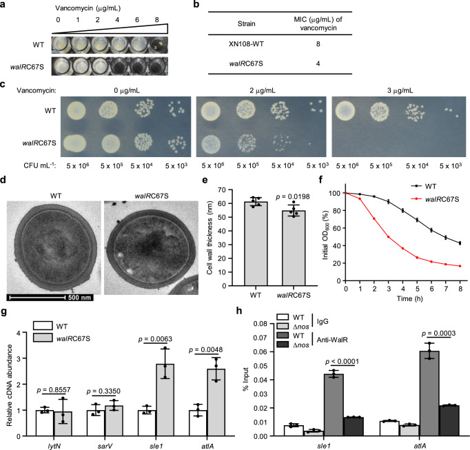
Treatment of Staphylococcus aureus infections is a constant challenge due to emerging resistance to vancomycin, a last-resort drug. S-nitrosylation, the covalent attachment of a nitric oxide (NO) group to a cysteine thiol, mediates redox-based signaling for eukaryotic cellular functions. However, its role in bacteria is largely unknown. Here, proteomic analysis revealed that S-nitrosylation is a prominent growth feature of vancomycin-intermediate S. aureus. Deletion of NO synthase (NOS) or removal of S-nitrosylation from the redox-sensitive regulator MgrA or WalR resulted in thinner cell walls and increased vancomycin susceptibility, which was due to attenuated promoter binding and released repression of genes involved in cell wall metabolism. These genes failed to respond to H2O2-induced oxidation, suggesting distinct transcriptional responses to alternative modifications of the cysteine residue. Furthermore, treatment with a NOS inhibitor significantly decreased vancomycin resistance in S. aureus. This study reveals that transcriptional regulation via S-nitrosylation underlies a mechanism for NO-mediated bacterial antibiotic resistance.


Conflict of interest statement

The authors declare no competing interests.

Figures

Fig. 1
Fig. 1. Identification of the transcription factor MgrA as a target of S-nitrosylation.
a Summary of MS identified NO-modified proteins in S. aureus categorized by biological pathway. b Transcriptional regulators identified to be S-nitrosylated are listed. c Higher-energy collision dissociation mass spectrum of the cystine 12-containing peptide from MgrA. S-nitrosylated cysteine was labeled with isobaric iodoTMT and marked with io. The cysteine residue Cys12 of MgrA is located within the MarR-type HTH domain (d) and located at the interface of MgrA dimer (e). f S-nitrosylation of MgrA purified from the XN108 WT or Δnos strain was detected by western blot. S-nitrosylated MgrA (MgrA-SNO) was detected with an anti-TMT antibody. Total MgrA (MgrA-His) was detected with an anti-His antibody. Data are representative of n = 3 biological replicates. Source data are provided as a Source Data file.
Fig. 2
Fig. 2. NO-mediated MgrA S-nitrosylation promotes the resistance of VISA to vancomycin.
a MICs were assessed after the WT S. aureus XN108, mgrAC12S mutant and Δnos mutant strains were grown under vancomycin conditions in a 96-well plate with shaking at 200 rpm for 48 h at 37 °C. Data are representative of n = 3 biological replicates. b The growth of all the strains on normal condition or medium with different concentrations of vancomycin as indicated on agar plates was measured by a plate-sensitivity assay. Data are representative of n = 3 biological replicates. CFU colony-forming units. Growth curve of the WT VISA strain XN108 (c) or Mu50 (d) in normal MH II medium (V-0 L-0) or medium containing 2 μg/mL vancomycin (V-2 L-0) or supplemented with 40 mM L-NAME (V-2 L-40) in 96-well plates with an initial OD600 of 0.001. These cultures were incubated at 37 °C without shaking overnight (13 h) and then grown with shaking at 200 rpm for 12 h. Data are means of n = 3 biological replicates with SD. Two-sided two-way ANOVA, ***p ≤ 0.001; ****p ≤ 0.0001. Source data are provided as a Source Data file.
Fig. 3
Fig. 3. NO is required for S-nitrosylation on MgrA to facilitate vancomycin resistance.
The WT and Δnos strains (ac) or the WT and mgrAC12S strains (df) were grown in MH II medium containing 2 μg/mL vancomycin in 96-well plates with shaking at 200 rpm for 48 h at 37 °C. OD600 was monitored to evaluate the growth when different concentrations of SNP were added. N-0, N-1, N-5 or N-20 represents 0, 1, 5 or 20 μM SNP respectively. Data are means of n = 3 biological replicates with SD. Two-sided two-way ANOVA, *p ≤ 0.05; **p ≤ 0.01; ***p ≤ 0.001; ****p ≤ 0.0001; ns not significant. Source data are provided as a Source Data file.
Fig. 4
Fig. 4. Redox-signal transduction by MgrA to vancomycin resistance is achieved by regulating cell wall thickness and cell autolysis.
a, b Cell wall thickness was measured by transmission electron microscopy (TEM). Representative TEM images of the WT, mgrAC12S and Δnos strains cultured with shaking at 220 rpm for 16 h at 37 °C are displayed (a). Scale bar = 500 nm. Cell wall thickness of n = 5 individual cells from each strain were measured three times randomly and expressed as means with SD (b). Triton X-100-induced autolysis of the WT and Δnos strains (c) or the WT and mgrAC12S strains (d) was measured in Tri-HCl buffer supplemented with or without 5 μM SNP (displayed as N-5 or N-0). The percentage of the initial OD600 was displayed. Data are means of n = 3 biological replicates with SD. e The transcriptional levels of the indicated genes that reported to regulate autolysis activity were tested in the WT and mgrAC12S mutant strains by using qRT-PCR. Data are means of n = 3 biological replicates with SD. The transcriptional levels of lytN and sarV were measured in both strains after incubation with different concentrations of SNP (f) or H2O2 (g) as indicated. Data are means of n = 3 biological replicates with SD. Two-sided unpaired Student’s t-test, *p ≤ 0.05; ***p ≤ 0.001; ****p ≤ 0.0001; ns not significant (b, e, f, g). Source data are provided as a Source Data file.
Fig. 5
Fig. 5. Removal of MgrA S-nitrosylation impairs its DNA binding activity.
ChIP-qPCR was used to evaluate the occupancy of endogenous WT MgrA or MgrAC12S at the promoters of lytN and sarV (a, b), and the occupancy of endogenous WT MgrA in the WT or Δnos strains (c, d). Data are means of n = 3 biological replicates with SD. Two-sided unpaired Student’s t-test, ****p ≤ 0.0001. DNA binding efficacy of WT and C12S mutant MgrA at lytN (e) or sarV (f) promoters was compared by using gel-shift assay. Different concentrations of purified MgrA or MgrAC12S protein were incubated with a FAM-labeled probe containing the MgrA binding sequence on the lytN or sarV promoter region. The probe concentrations were indicated as plytN or psarV. The upper bands represent probes bound with protein; the lower bands represent free probes. Data are representative of n = 3 biological replicates. Source data are provided as a Source Data file.
Fig. 6
Fig. 6. WalR adopts S-nitrosylation to confer vancomycin resistance.
a MICs were assessed after the WT and walRC67S mutant strains were grown under vancomycin conditions in a 96-well plate with shaking at 200 rpm for 48 h at 37 °C. Data are representative of n = 3 biological replicates. b The difference in MICs observed in a is displayed. c The growth of both strains on normal condition or medium containing different concentrations of vancomycin as indicated on agar plates was measured by a plate-sensitivity assay. CFU colony-forming units. d, e Cell wall thickness was measured by transmission electron microscopy (TEM). Representative TEM images of the WT and walRC67S strains cultured with shaking at 220 rpm for 16 h at 37 °C are displayed (d). Scale bar = 500 nm. Cell wall thickness of n = 5 individual cells from each strain were measured three times randomly and expressed as means with SD (e). f Triton X-100-induced autolysis of the WT and walRC67S mutant strains was measured in 96-well plate with shaking at 200 rpm for 8 h at 37 °C. The percentage of the initial OD600 was displayed. Data are means of n = 3 biological replicates with SD. g The transcriptional levels of selected genes that were reported to regulate autolysis were tested in all the strains by using qRT-PCR. Data are means of n = 3 biological replicates with SD. h ChIP-qPCR was used to evaluate the occupancy of WT WalR or WalRC67S at promoters of sle1 and altA in the WT and Δnos strains. Data are means of n = 3 biological replicates with SD. Two-sided unpaired Student’s t-test, *p ≤ 0.05; **p ≤ 0.01; ***p ≤ 0.001; ****p ≤ 0.0001; ns not significant (e, g, h). Source data are provided as a Source Data file.
Fig. 7
Fig. 7. Mechanism of S-nitrosylation signal transduction to vancomycin resistance.
S. aureus has evolved saNOS to generate NO. When the NO signal is on, MgrA and WalR undergo S-nitrosylation on the cysteine thiol, thereby initiating the transcriptional repression signals to the downstream genes such as lytN and sarV to inhibit autolysin expression, which leads to a thickened cell wall and increased resistance to vancomycin. When the NO signal is off, the autolysin production will be increased, thus resulting in accelerated cell wall degradation and increased susceptibility to vancomycin. The lytN gene encodes an autolysin protein. The sarV gene encodes a SarV protein, which is a positive regulator of autolysin genes. GlcNAc, N-acetylglucosamine. MurNAc, N-acetylmuramic acid. Pentapeptide, L-Ala–D-Gln–L-Lys–D-Ala–D-Ala.

Similar articles

Cited by

References

    1. Tong SY, Davis JS, Eichenberger E, Holland TL, Fowler VG., Jr. Staphylococcus aureus infections: epidemiology, pathophysiology, clinical manifestations, and management. Clin. Microbiol. Rev. 2015;28:603–661. - PMC - PubMed
    1. Howden BP, Davies JK, Johnson PD, Stinear TP, Grayson ML. Reduced vancomycin susceptibility in Staphylococcus aureus, including vancomycin-intermediate and heterogeneous vancomycin-intermediate strains: resistance mechanisms, laboratory detection, and clinical implications. Clin. Microbiol. Rev. 2010;23:99–139. - PMC - PubMed
    1. Liu J, Gefen O, Ronin I, Bar-Meir M, Balaban NQ. Effect of tolerance on the evolution of antibiotic resistance under drug combinations. Science. 2020;367:200–204. - PubMed
    1. Jernigan JA, et al. Multidrug-resistant bacterial infections in U.S. hospitalized patients, 2012-2017. N. Engl. J. Med. 2020;382:1309–1319. - PMC - PubMed
    1. Yang X, et al. Clonal and drug resistance dynamics of methicillin-resistant Staphylococcus aureus in pediatric populations in China. Pediatr. Investig. 2019;3:72–80. - PMC - PubMed

Publication types

MeSH terms