

Various effects of 11,12 EET rescue wound healing in a combined model of diabetes and ischemia
- PMID: 37085527
- PMCID: PMC10121596
- DOI: 10.1038/s41598-023-33400-y
Various effects of 11,12 EET rescue wound healing in a combined model of diabetes and ischemia
Abstract
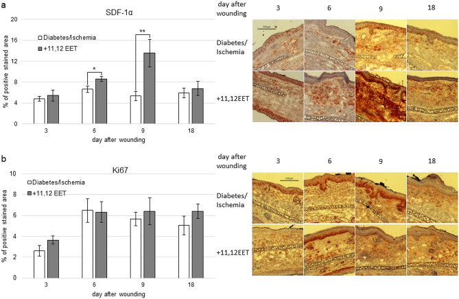
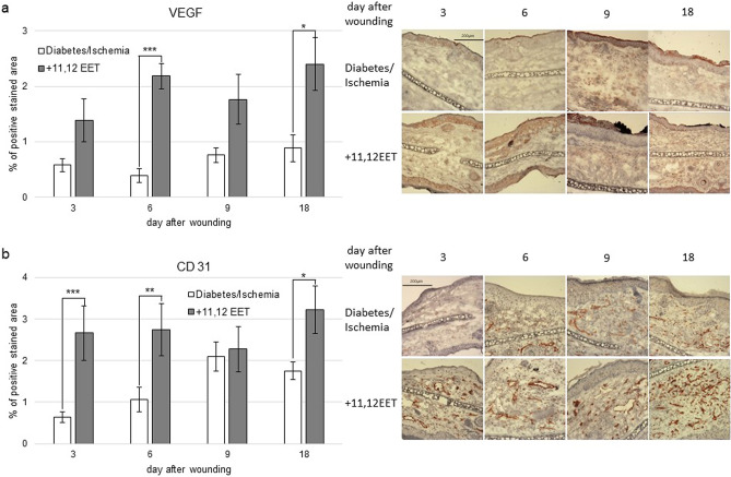
Chronic non healing wounds in diabetic patients still impose a major problem in modern medicine. Especially additional peripheral vascular disease complicates treatment success in these patients. Thus, we analyzed the effects of 11,12 epoxyeicosatrienoic acid (EET) in a combined model of hyperglycemia and ischemia in mice. Hyperglycemia was induced by Streptozotozin 2 weeks prior to wounding. 3 days before wound creation 2 of the 3 suppling vessels of the moue ear were cautherized for ischemia. Either 11,12 EET or solvent for control was applied. Wound closure as well as TNF-α, TGF-β, SDF-1α, VEGF, CD31, and Ki67 were measured. The wounds closed on day 14.4 ± 0.4 standard deviation (SD). 11,12 EET treatment enhanced healing to 9.8 ± 0.6 SD. TNF-α level was augmented on day 9 compared to control and receded on day 18. TGF-β seemed to be elevated all days observed after 11,12 EET treatment. SDF-1α was enhanced on day 6 and 9 by 11,12 EET, and VEGF on day 6 and 18 as well as CD13 on day 3, 6, and 18. 11,12 EET did not alter Ki67. 11,12 EET are able to rescue deteriorated wound healing in a combined model of hyperglycamia and ischemia by resolution of inflammation, augmentation of neovascularization and increasing expression of TGF-β as well as SDF-1α.
© 2023. The Author(s).
Conflict of interest statement
The authors declare no competing interests.
Figures


Similar articles
-
11,12 and 14,15 epoxyeicosatrienoic acid rescue deteriorated wound healing in ischemia.PLoS One. 2019 Jan 16;14(1):e0209158. doi: 10.1371/journal.pone.0209158. eCollection 2019. PLoS One. 2019. PMID: 30650075 Free PMC article.
-
11,12 Epoxyeicosatrienoic Acid Rescues Deteriorated Wound Healing in Diabetes.Int J Mol Sci. 2021 Oct 28;22(21):11664. doi: 10.3390/ijms222111664. Int J Mol Sci. 2021. PMID: 34769092 Free PMC article.
-
Lentiviral gene transfer of SDF-1alpha to wounds improves diabetic wound healing.J Surg Res. 2007 Nov;143(1):35-42. doi: 10.1016/j.jss.2007.03.051. J Surg Res. 2007. PMID: 17950070
-
Effect of genipin crosslinked chitosan scaffolds containing SDF-1 on wound healing in a rat model.Mater Sci Eng C Mater Biol Appl. 2020 Apr;109:110368. doi: 10.1016/j.msec.2019.110368. Epub 2019 Dec 6. Mater Sci Eng C Mater Biol Appl. 2020. PMID: 32228920
-
[The modern approach to wound treatment].Med Pregl. 2000 Jul-Aug;53(7-8):363-8. Med Pregl. 2000. PMID: 11214479 Review. Croatian.
Cited by
-
Severity-Dependent Long-Term Post-Traumatic Changes in the Circulating Oxylipin Profile.Int J Mol Sci. 2024 Dec 17;25(24):13530. doi: 10.3390/ijms252413530. Int J Mol Sci. 2024. PMID: 39769293 Free PMC article.
References
-
- Nguyen KT, et al. Deficient cytokine expression and neutrophil oxidative burst contribute to impaired cutaneous wound healing in diabetic, biofilm-containing chronic wounds. Wound Repair Regen. Off. Publ. Wound Heal. Soc. Eur. Tissue Repair Soc. 2013;21:833–841. doi: 10.1111/wrr.12109. - DOI - PubMed
Publication types
MeSH terms
Substances
LinkOut - more resources
Full Text Sources
Other Literature Sources
Medical
Miscellaneous
