. 2023 Apr 21;13(1):6555.
doi: 10.1038/s41598-023-33543-y.

Degeneration of muscle spindles in a murine model of Pompe disease

Affiliations

Degeneration of muscle spindles in a murine model of Pompe disease

Bridgette Watkins et al. Sci Rep. .

Erratum in

Abstract

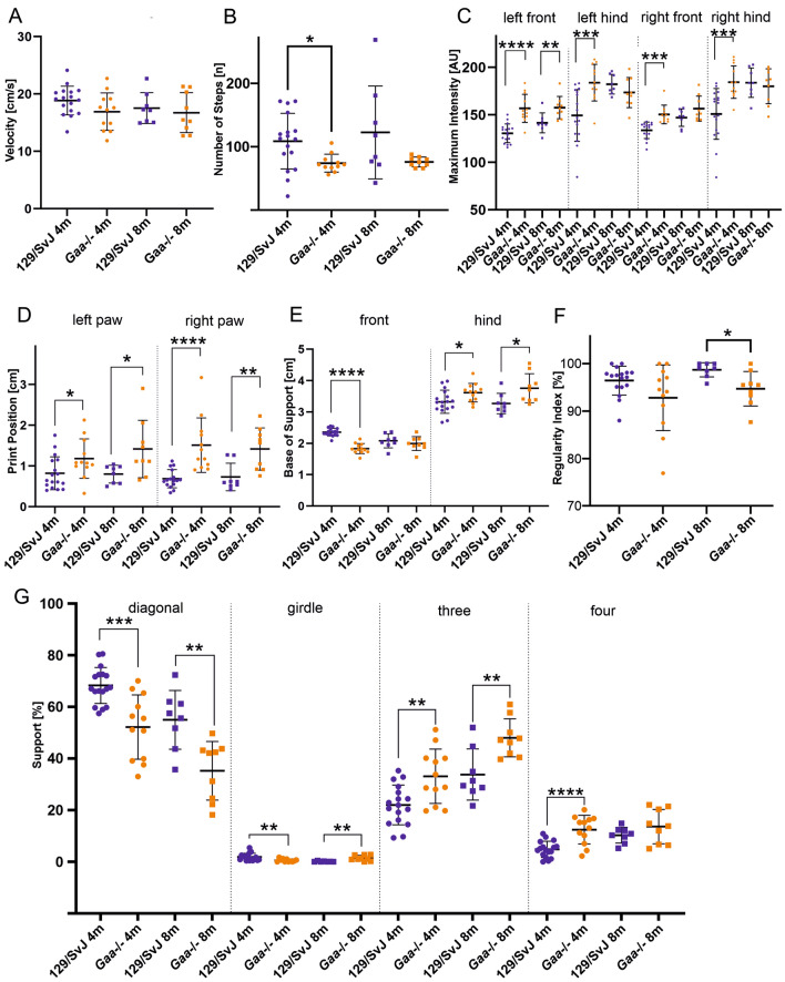
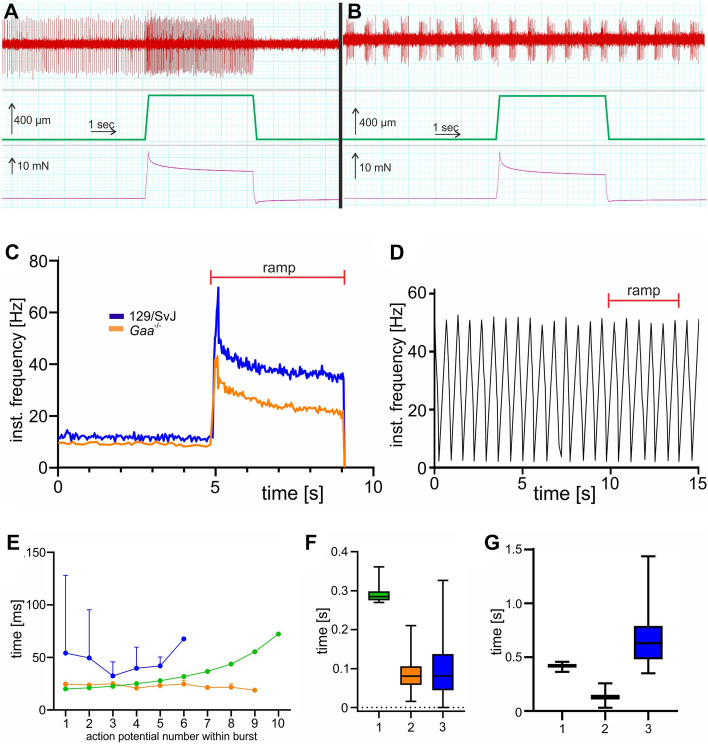
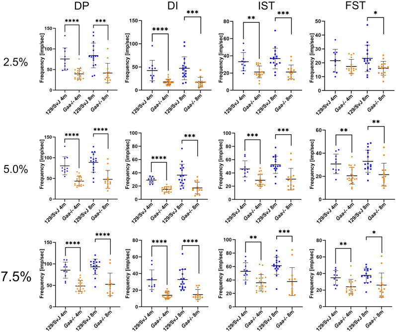
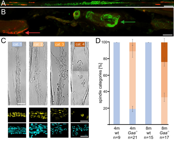
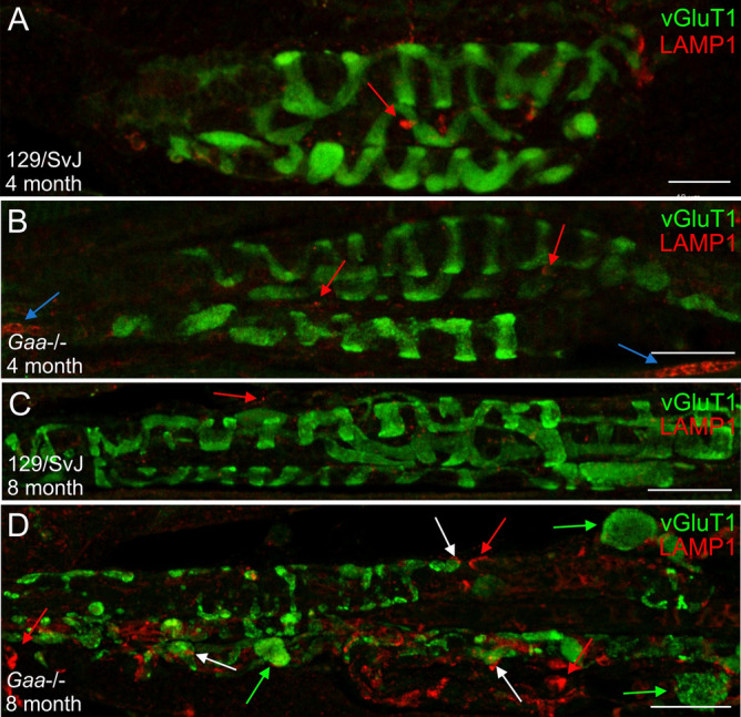
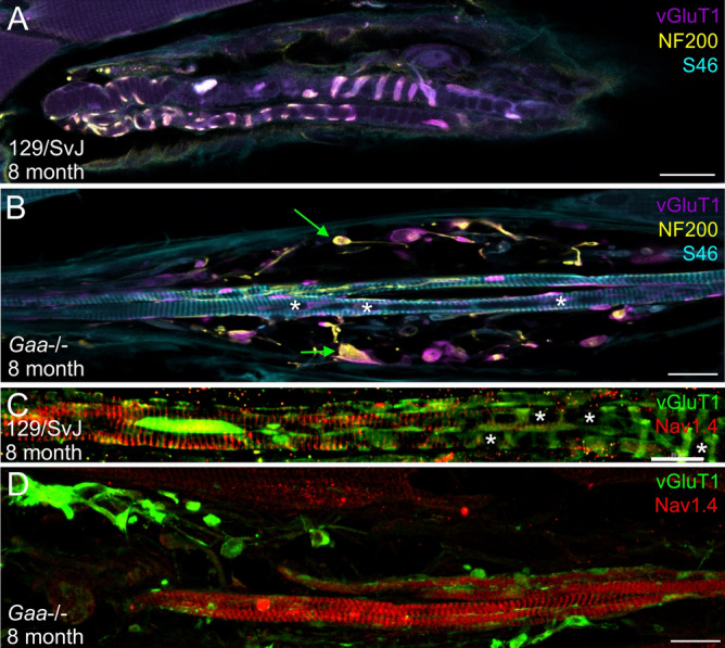
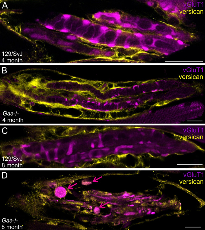
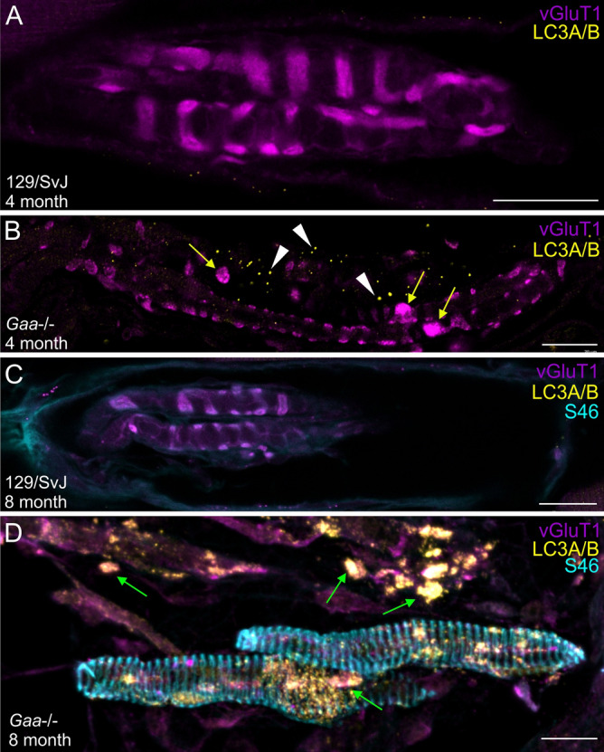
Pompe disease is a debilitating medical condition caused by a functional deficiency of lysosomal acid alpha-glucosidase (GAA). In addition to muscle weakness, people living with Pompe disease experience motor coordination deficits including an instable gait and posture. We reasoned that an impaired muscle spindle function might contribute to these deficiencies and therefore analyzed proprioception as well as muscle spindle structure and function in 4- and 8-month-old Gaa-/- mice. Gait analyses showed a reduced inter-limb and inter-paw coordination in Gaa-/- mice. Electrophysiological analyses of single-unit muscle spindle proprioceptive afferents revealed an impaired sensitivity of the dynamic and static component of the stretch response. Finally, a progressive degeneration of the sensory neuron and of the intrafusal fibers was detectable in Gaa-/- mice. We observed an increased abundance and size of lysosomes, a fragmentation of the inner and outer connective tissue capsule and a buildup of autophagic vacuoles in muscle spindles from 8-month-old Gaa-/- mice, indicating lysosomal defects and an impaired autophagocytosis. These results demonstrate a structural and functional degeneration of muscle spindles and an altered motor coordination in Gaa-/- mice. Similar changes could contribute to the impaired motor coordination in patients living with Pompe disease.


Conflict of interest statement

The authors declare no competing interests.

Figures

Figure 1
Figure 1
Gaa−/− mice have an abnormal motor coordination. Automatic gait analysis revealed that many general locomotor parameters were similar in wildtype 129/SvJ mice (blue dots) and Gaa−/− mice (orange dots), including velocity of movement (A) and number of steps (B; the slightly reduced number of steps in 4-month-old mice is most likely due to the weight difference). Other parameters are different between Gaa−/− mice and age-matched control mice due to their different muscle force, including the maximum intensity of the footprints (C). On the other hand, both mouse lines behaved differently with respect to locomotion coordination parameters, including the print position (the distance between the position of the hind paw and the position of the previously placed front paw on the same side of the body and in the same step cycle; D), the base of support for the front- and hind limbs (E), as well as the regularity index (F). Moreover, the time the mice were supported by contacting the ground with the diagonal and girdle sides limbs as well as the time the animal was supported by three or four limbs was longer in Gaa−/− mice compared to 129/SvJ control mice (G). For a complete list of gait parameters analyzed see Supplementary Table 1. The bars show the mean ± SD with N = 17 (4-month-old 129SvJ) and N = 12 (4-month-old Gaa−/−), N = 8 (8-month-old 129SvJ), N = 9 (8-month old Gaa−/−) mice. Statistical significance was calculated using the unpaired student’s t-test.
Figure 2
Figure 2
The response to stretch in muscle spindles from Gaa−/− mice is impaired. A representative example for the response of muscle spindle afferents to a ramp-and-hold stretch from 129/SvJ wildtype mice is shown in panel (A). Muscle spindles from wildtype mice had a constant resting discharge of approximately 10 Hz and responded to stretch with an increase in their instantaneous frequency (A). In contrast, 70% of the muscle spindles from 8-month-old Gaa−/− mice (orange line in panel (C)) responded to stretch, but had a lower frequency at all time points during a stretch compared to age-matched 129/SvJ wildtype mice. Approximately 30% of the 8-month-old Gaa−/− mice fired bursts at rest and did not respond to stretch (B). In some spindles, the bursts were rather regular even during a ramp-and-hold stretch (D). In other muscle spindles from 8-month-old Gaa−/− mice, which showed the bursting behavior at rest, the bursts varied with respect to the action potential frequency during the bursts (E), the duration of the bursts (F) and the interburst interval (duration of the silent period; (G)). Three representative spindles from three different 8-month-old Gaa−/− mice are shown to illustrate the spectrum of the busts. The red bars in panels (C,D) indicate the duration of the ramp-and-hold stretch. The middle parts of panels (A,B) show the length change (5% of L0) and the lower parts show the passive tension generated by the muscle in response to the stretch. No difference between wildtype and Gaa−/− mice was observed with respect to the passive tension generated in response to the length change.
Figure 3
Figure 3
Stretch-responsive muscle spindles from Gaa−/− mice have lower instantaneous frequencies compared to control mice at all time points during ramp-and-hold stretches. The frequency at four time points during a ramp-and-hold stretch (dynamic peak (DP), dynamic index (DI), initial static time (IST) and final static time (FST); for details of these time points see “Methods” section) were lower in Gaa−/− mice (orange dots) compared to age-matched wildtype 129/SvJ control mice (blue dots). This was independent of the length change (2.5, 5.0 and 7.5% of L0) and of the age of the mice (4- and 8-month-old mice, respectively). Each dot represents the recording of a single muscle spindle. Bars show the mean ± SD with N = 4 (4-month-old 129SvJ), N = 8 (4-month-old Gaa−/−), N = 6 (8-month-old 129SvJ) and N = 16 (8-month-old Gaa−/−). Statistical significance was calculated using the unpaired student t-test.
Figure 4
Figure 4
Quantification of the degenerative changes in muscle spindles from Gaa−/− mice. Intrafusal fibers are innervated by sensory nerve terminals in the central (equatorial) region (stained with antibodies against vGluT1; green channel in (A,B)) and by γ-motoneurons in the polar regions, which form a cholinergic synapse (indicated by α-bungarotoxin labeling of the AChRs; red channel in (A,B)). Note the normal structure of the intrafusal fiber innervation in muscle spindles from 8-month-old 129/SvJ mice (A) and the severely degenerated innervation in the 8-month-old Gaa−/− mice ((B); corresponding to a category 4 muscle spindle, see below). The sensory nerve terminal has retracted from the intrafusal fiber and has formed a large varicosity (green arrow). AChRs have disaggregated (red arrow) and the intrafusal fiber has lost its elongated shape and formed a spherical myoball-like structure. Panel (C) shows representative examples of the four different categories used to characterize the different levels of degeneration. For a more detailed description of the categories, see “Methods” section. The lower two rows of panel (C) show representative examples corresponding to the different categories of the morphology of the sensory ending (green channel) and the distribution of the nuclei (blue channel), respectively. Morphological analysis of 4- and 8-month-old muscle spindles from wildtype (wt) and Gaa−/− mice revealed a progressive increase of the number of damaged muscle spindles demonstrating the progressive degenerative changes (panel (D)). Bars show the mean ± SD with N = 3; n represents the number of muscle spindles analyzed. Color-coding of the different categories is identical in panels (C,D). Scale bar: (A) 50 µm, (B) 20 µm, (C) 20 µm.
Figure 5
Figure 5
Accumulation and aggregation of lysosomes in muscle spindles from Gaa−/− mice. The structure and distribution of lysosomes was investigated using anti-LAMP1 antibodies in 4-month-old (A,B) and 8-month-old (C,D) 129/SvJ control mice (A,C) and Gaa−/− mice (B,D). The structure of the sensory nerve terminal (stained by antibodies against vGluT1; green channel) was indistinguishable in 4- and 8-month-old muscle spindles from wildtype mice and in 4-month-old Gaa−/− mice. In contrast, in 8-month-old Gaa−/− mice (category 2), the sensory nerve terminal had formed several varicosities within the spindle matrix (green arrows in panel (D)), had retraced from the intrafusal fibers and had lost the typical annulospiral morphology. Lysosomes were barely detectable in sections from 129/SvJ mice at 4- and 8 months of age (red arrows in panels (A,C)). In contrast, lysosomes were significantly enlarged and had formed aggregates in 4-month-old Gaa−/− mice (red and blue arrows in panel (B), respectively). By 8 months (panel (D)), lysosomes were abundant, highly aggregated (red arrows) and often associated with the varicosities formed by the sensory nerve terminals of Gaa−/− mice (white arrows). Scale bars: 20 µm.
Figure 6
Figure 6
The varicosities formed by the sensory nerve terminal contain cytoskeletal elements and proteins of the polar region redistribute into the central region of intrafusal fiber’s in 8-month-old Gaa−/− mice. Muscle spindles from 8-month-old wildtype (A,C) and Gaa−/− (B,D) mice were stained with antibodies against vGluT1 (purple channel in panels A and B and green channel in panels (C,D)), neurofilament 200 (NF 200; yellow channel in (A,B)), the myosin heavy chain 6 (S46 antibody; blue channel in (A,B)) and the voltage-gated sodium channel Nav1.4 (red channel in (C,D)). In muscle spindles from wildtype 129/SvJ mice, neurofilament 200 immunoreactivity codistributed with the sensory nerve terminal (labeled by the anti-vGluT1 antibodies). The typical annulospiral morphology of the nerve terminal is detectable. In contrast, in muscle spindles from age-matched Gaa−/− mice (panels (B,D) show category 3 muscle spindles), the contact between the sensory nerve terminal and the intrafusal fibers was lost and the sensory nerve terminals had retracted to form numerous varicosities. The varicosities labeled by vGluT1 contained neurofilament 200 immunoreactivity (green arrows in panel (B)), demonstrating the accumulation of cytoskeletal elements in these structures. Note the absence of labeling with the S46 antibody in the central region of 8-month-old wildtype intrafusal fibers (A) and its presence in the central region of intrafusal fibers from age-matched Gaa−/− mice (B). Likewise, the voltage-gated sodium channel Nav1.4 was concentrated underneath the subsarcolemmal plasma membrane in the central region of intrafusal fibers of wildtype animals (C) whereas it was present throughout the intrafusal fiber in 8-month-old Gaa−/− mice (D). Asterisks in panels (B,C) indicate the central region of intrafusal fibers. Scale bars: 20 µm.
Figure 7
Figure 7
The muscle spindle connective tissue capsule deteriorates in 8-month-old Gaa−/− mice. Muscle spindles from 4- (A,B) and 8-month-old (C,D) wildtype (A,C) and Gaa−/− (B,D) mice were stained with antibodies against vGluT1 (purple channel) and the extracellular matrix protein versican (yellow channel). The antibodies labeled the inner and outer capsule of muscle spindles in wildtype and 4-month-old Gaa−/− mice. In 8-month-old Gaa−/− mice, the labeling was distributed throughout the muscle spindle and the inner- and outer capsule appeared fragmented. Note the vGluT1-positive varicosities (purple arrows in panel (D)) and the punctate versican immunoreactivity in the 8-month-old Gaa−/− mice. Scale bar: 20 µm.
Figure 8
Figure 8
Autophagosomes accumulate in muscle spindles from Gaa−/− mice. Muscle spindles from 4- (A,B) and 8-month-old (C,D) wildtype (A,C) and Gaa−/− (B,D) mice were stained with antibodies against vGluT1 (purple channel) and the autophagosome marker LC3A/B (yellow channel). Muscle spindles from 8-month-old wildtype and Gaa−/− mice were additionally stained with antibodies against myosin heavy chain 6 (S46 antibody; blue channel in panels (C,D)). While autophagosomes were mostly absent in muscle spindles from 4- and 8-month-old wildtype mice, they appeared in 4-month-old Gaa−/− mice (white arrowheads in panel (B); category 2 muscle spindle). In 8-month-old Gaa−/− mice autophagosomes were abundant (arrows in panel (D); category 3 muscle spindle) and the LC3A/B immunoreactivity was associated with intrafusal fibers (arrows in panel (D)) and with the varicosities formed by the sensory nerve terminal (green arrows in panel (D)). Note the formation of small varicosities by the sensory nerve terminal in muscle spindles from 4-month-old Gaa−/− mice (yellow arrows in panel (B)). S46 immunoreactivity was absent in the central region of intrafusal fibers of 8-month-old 129/SvJ mice (C), but present in age-matched Gaa−/− mice (D), confirming the reorganization of the intrafusal contractile filaments. Scale bars: 20 µm.

Similar articles

Cited by

References

    1. van der Ploeg AT, Reuser AJ. Pompe's disease. Lancet. 2008;372:1342–1353. doi: 10.1016/S0140-6736(08)61555-X. - DOI - PubMed
    1. Lim JA, Li L, Raben N. Pompe disease: From pathophysiology to therapy and back again. Front. Aging Neurosci. 2014;6:177. doi: 10.3389/fnagi.2014.00177. - DOI - PMC - PubMed
    1. Kohler L, Puertollano R, Raben N. Pompe disease: From basic science to therapy. Neurotherapeutics. 2018;15:928–942. doi: 10.1007/s13311-018-0655-y. - DOI - PMC - PubMed
    1. Peruzzo P, Pavan E, Dardis A. Molecular genetics of Pompe disease: A comprehensive overview. Ann. Transl. Med. 2019;7:278. doi: 10.21037/atm.2019.04.13. - DOI - PMC - PubMed
    1. Martiniuk F, Bodkin M, Tzall S, Hirschhorn R. Isolation and partial characterization of the structural gene for human acid alpha glucosidase. DNA Cell Biol. 1991;10:283–292. doi: 10.1089/dna.1991.10.283. - DOI - PubMed

Publication types

Substances