

3-D and 2-D reconstruction of bladders for the assessment of inter-session detection of tissue changes: a proof of concept
- PMID: 37085675
- PMCID: PMC10497453
- DOI: 10.1007/s11548-023-02900-7
3-D and 2-D reconstruction of bladders for the assessment of inter-session detection of tissue changes: a proof of concept
Abstract
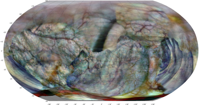
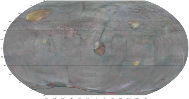
Purpose: Abnormalities in the bladder wall require careful investigation regarding type, spatial position and invasiveness. Construction of a 3-D model of the bladder is helpful to ensure adequate coverage of the scanning procedure, quantitative comparison of bladder wall textures between successive sessions and finding back previously discovered abnormalities.
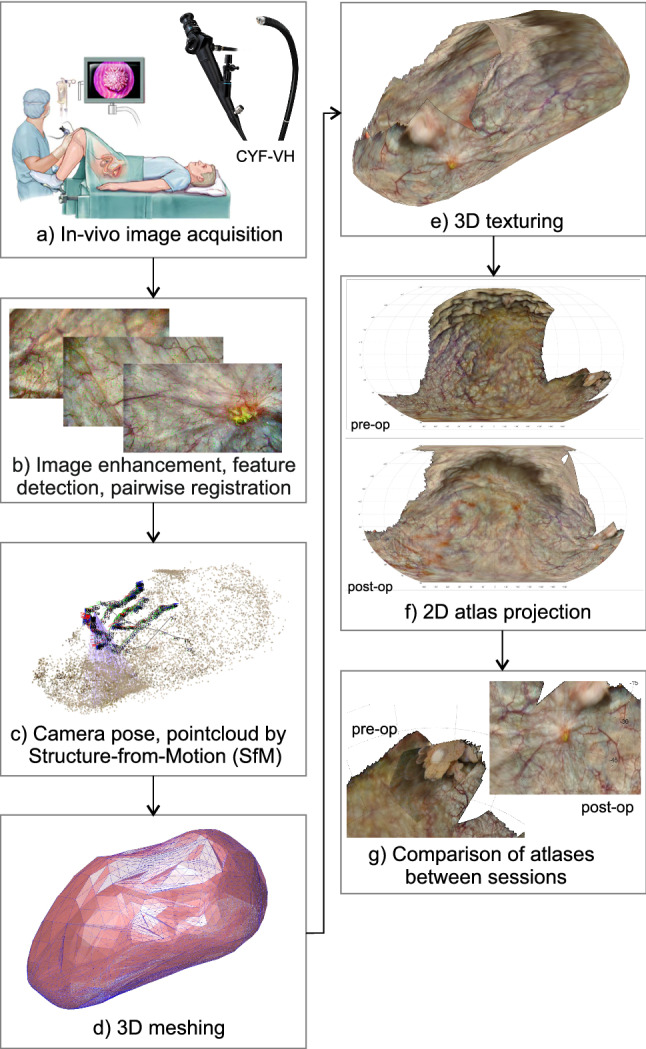
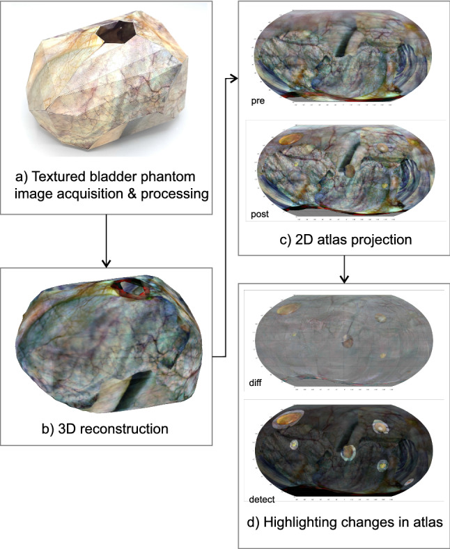
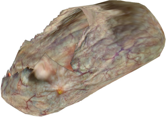
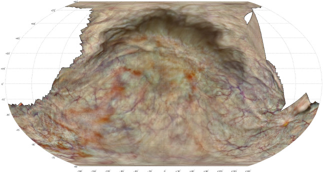
Methods: Videos of both an in vivo bladder and a textured bladder phantom were acquired. Structure-from-motion and bundle adjustment algorithms were used to construct a 3-D point cloud, approximate it by a surface mesh, texture it with the back-projected camera frames and draw the corresponding 2-D atlas. Reconstructions of successive sessions were compared; those of the bladder phantom were co-registered, transformed using 3-D thin plate splines and post-processed to highlight significant changes in texture.
Results: The reconstruction algorithms of the presented workflow were able to construct 3-D models and corresponding 2-D atlas of both the in vivo bladder and the bladder phantom. For the in vivo bladder the portion of the reconstructed surface area was 58% and 79% for the pre- and post-operative scan, respectively. For the bladder phantom the full surface was reconstructed and the mean reprojection error was 0.081 mm (range 0-0.79 mm). In inter-session comparison the changes in texture were correctly indicated for all six locations.
Conclusion: The proposed proof of concept was able to perform 3-D and 2-D reconstruction of an in vivo bladder wall based on a set of monocular images. In a phantom study the computer vision algorithms were also effective in co-registering reconstructions of successive sessions and highlighting texture changes between sessions. These techniques may be useful for detecting, monitoring and revisiting suspicious lesions.
Keywords: 3-D reconstruction; Bladder; Computer vision; Monocular; Proof of concept.
© 2023. The Author(s).
Conflict of interest statement
The authors declare that they have no conflict of interest.
Figures


References
-
- Brierley J, Gospodarowicz M, Wittekind C. TNM classification of malignant tumors. 8. Hoboken: Wiley; 2017.
-
- Bertero L, Massa F, Metovic J, Zanetti R, Castellano I, Ricardi U, Papotti M, Cassoni P (2018) Eighth edition of the UICC classification of malignant tumours: An overview of the changes in the pathological TNM classification criteria-What has changed and why? Springer. 10.1007/s00428-017-2276-y - PubMed
-
- Holmäng S, Johansson SL (2002) Stage Ta-T1 bladder cancer: the relationship between findings at first follow up cystoscopy and subsequent recurrence and progression. 10.1016/S0022-5347(05)65168-3 - PubMed
MeSH terms
Grants and funding
LinkOut - more resources
Full Text Sources
