

Thygeson's superficial punctate keratitis in children
- PMID: 37085721
- PMCID: PMC10630495
- DOI: 10.1038/s41433-023-02533-9
Thygeson's superficial punctate keratitis in children
Abstract
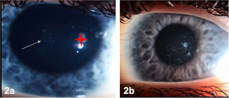
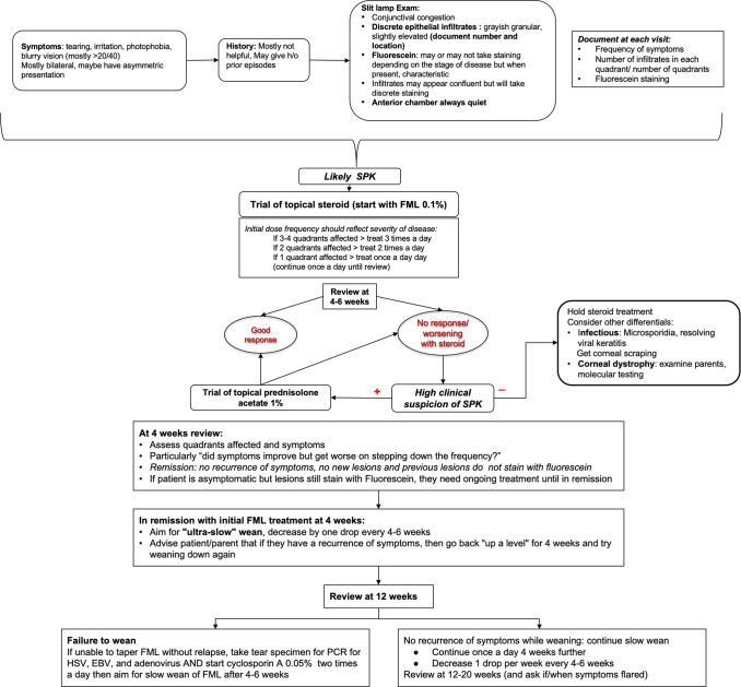
Objective: To describe the first paediatric case series of Thygesons' superficial punctate keratitis (TSPK) with management outcomes.
Methods: A retrospective chart review was done for all children either diagnosed at initial presentation or referred with TSPK from 01/2012 to 08/2021 at a tertiary children's hospital. Records were assessed for signs, symptoms, diagnosis, steroid and cyclosporine 0.05% use. The main outcome measures were visual acuity, treatment response and total steroid exposure.
Results: Fifteen children (7 females), mean age at presentation 8 ± 4 years were included. All had bilateral disease and a BCVA of >20/40 in the better eye. All patients received topical fluorometholone 0.1%, (FML) initially. 80% had a good response to FML. Corneal scraping was done to exclude infectious causes in four cases due to poor initial response or clinical suspicion. All 4 needed EUA for scraping and anterior segment OCT, after which 2 had molecularly confirmed TGFBI-related stromal dystrophy. For the rest, slow steroid taper was done every 4-6 weeks and recurrences were treated by increasing steroid frequency. Cyclosporine 0.05% was started in nine patients (69%), 8 ± 6 months after initial presentation. The decrease in total steroid exposure per week after starting cyclosporine was statistically significant (p < 0.05).
Conclusion: Children with TSPK respond quickly to steroids, however, recurrences are common, necessitating a slow taper. Non-response to steroid needs careful reconsideration of the diagnosis and may necessitate the use of an EUA. Using cyclosporine 0.05% reduces the total steroid exposure in TSPK.
© 2023. The Author(s), under exclusive licence to The Royal College of Ophthalmologists.
Conflict of interest statement
The authors declare no competing interests.
Figures


Comment on
-
Thygeson's superficial punctate keratitis (TSPK): a paediatric case report and review of the literature.BMC Ophthalmol. 2021 Jan 29;21(1):64. doi: 10.1186/s12886-020-01790-6. BMC Ophthalmol. 2021. PMID: 33514353 Free PMC article. Review.
References
-
- Chan TCY, Chau HHT, Bhat AK, Nischal KK, Jhanji V. Thygeson’s superficial punctate keratitis. J EuCornea. 2019;3:5–8. doi: 10.1016/j.xjec.2019.11.001. - DOI
Publication types
MeSH terms
Substances
Grants and funding
LinkOut - more resources
Full Text Sources
Miscellaneous
