

Rheumatism as a cause of cardiac hemangioma: a rare case report and review of literature with special focus on etiology
- PMID: 37085767
- PMCID: PMC10122334
- DOI: 10.1186/s12872-023-03241-8
Rheumatism as a cause of cardiac hemangioma: a rare case report and review of literature with special focus on etiology
Abstract
Background: Cardiac hemangioma is a very rare benign tumor of the heart which accounts for 1-2% of all primary cardiac tumors. Multiple cardiac hemangiomas are even rarer with only three cases published in the literature. Pathologically it can be divided into cavernous hemangioma, capillary hemangioma, arteriovenous hemangioma, mixed-type hemangioma, and so on. At present, the etiology of cardiac hemangioma is not completely clear. In this study, we present multiple cardiac hemangiomas located in the right atrium and discuss the new unreported possible cause (rheumatism) of cardiac hemangioma. This is the fourth case of multiple cardiac hemangiomas in the medical literature and the first time to present rheumatism as the cause of cardiac hemangioma.
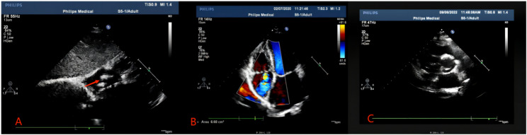
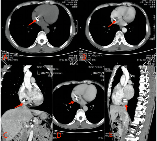
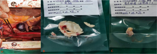
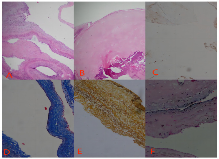
Case presentation: A 53-year-old man presented to the clinic with intermittent chest tightness and shortness of breath for 2 years. On echocardiography, multiple soft tissue masses in the right atrium were found. The patient had rheumatic heart disease with severe mitral stenosis and moderate tricuspid regurgitation. Two masses with a diameter of about 20 mm and 15 mm were seen in the right atrium. One mass was located on the inferior margin of the fossa ovalis and the other was adjacent to the inferior vena cava. Both masses were successfully removed surgically. The mitral valve replacement and tricuspid valve plasty were performed at the same time. The postoperative histopathology results confirmed the diagnosis of cavernous hemangioma.
Conclusion: The occurrence of multiple hemangiomas in the heart is possible, especially in the presence of rheumatism. Rheumatism is one of the possible etiologies of cardiac hemangioma. Cardiologists and cardiac surgeons should be aware of its occurrence and should consider cardiac hemangioma as a differential diagnosis especially in rheumatic heart disease patients when they present with soft tissue cardiac masses for accurate management.
Keywords: Benign heart tumors; Cardiac surgery; Hemangioma; Rheumatic heart disease; Rheumatism.
© 2023. The Author(s).
Conflict of interest statement
The authors have no competing interests to declare.
Figures
Similar articles
-
Super large cardiac hemangioma in right atrium and inferior vena cava: case report.J Cardiothorac Surg. 2019 Nov 5;14(1):186. doi: 10.1186/s13019-019-1016-6. J Cardiothorac Surg. 2019. PMID: 31690322 Free PMC article.
-
Diagnostic mystery-a rare right ventricular cardiac hemangioma: a case report.J Cardiothorac Surg. 2021 Dec 31;16(1):362. doi: 10.1186/s13019-021-01731-4. J Cardiothorac Surg. 2021. PMID: 34972529 Free PMC article.
-
Epicardial cardiac cavernous Haemangioma-a case report.BMC Cardiovasc Disord. 2019 Jul 29;19(1):179. doi: 10.1186/s12872-019-1156-6. BMC Cardiovasc Disord. 2019. PMID: 31357944 Free PMC article.
-
Cardiac autotransplantation for removal of left atrial hemangioma and a review of the literature.Heart Surg Forum. 2009 Oct;12(5):E279-84. doi: 10.1532/HSF98.20091027. Heart Surg Forum. 2009. PMID: 19833595 Review.
-
Cavernous hemangioma of the mitral valve: a case report and review of literature.J Cardiovasc Med (Hagerstown). 2009 May;10(5):420-2. doi: 10.2459/JCM.0b013e32832915a6. J Cardiovasc Med (Hagerstown). 2009. PMID: 19318977 Review.
Cited by
-
Cardiac interventricular septum hemangioma in a colon cancer patient treated with Capecitabine: A case report and review of literature.Clin Case Rep. 2024 Aug 19;12(8):e9331. doi: 10.1002/ccr3.9331. eCollection 2024 Aug. Clin Case Rep. 2024. PMID: 39161673 Free PMC article.
-
Mechanism of miR-503-5p on cardiac hemangioma and clinical validation.Am J Cancer Res. 2024 Nov 15;14(11):5304-5320. doi: 10.62347/EVMG4299. eCollection 2024. Am J Cancer Res. 2024. PMID: 39659925 Free PMC article.
-
Cardiac Tumors Causing Sudden Cardiac Death: A State-of-the-Art Review in Pathology.Cancers (Basel). 2025 Feb 17;17(4):669. doi: 10.3390/cancers17040669. Cancers (Basel). 2025. PMID: 40002264 Free PMC article. Review.
-
Mitral valve leaflet blood cyst treated with minimally invasive approach: a case report and review of literature.J Cardiothorac Surg. 2024 Jan 28;19(1):30. doi: 10.1186/s13019-024-02493-5. J Cardiothorac Surg. 2024. PMID: 38281941 Free PMC article. Review.
-
Cardiac Hemangiomas: A Five-Year Systematic Review of Diagnosis, Treatment, and Outcomes.Cancers (Basel). 2025 Apr 30;17(9):1532. doi: 10.3390/cancers17091532. Cancers (Basel). 2025. PMID: 40361457 Free PMC article. Review.
References
Publication types
MeSH terms
LinkOut - more resources
Full Text Sources
Medical
