Review
. 2023 Apr 21;18(1):27.
doi: 10.1186/s13024-023-00617-4.

Proteostasis failure exacerbates neuronal circuit dysfunction and sleep impairments in Alzheimer's disease

Affiliations
Review

Proteostasis failure exacerbates neuronal circuit dysfunction and sleep impairments in Alzheimer's disease

Christopher Daniel Morrone et al. Mol Neurodegener. .

Abstract

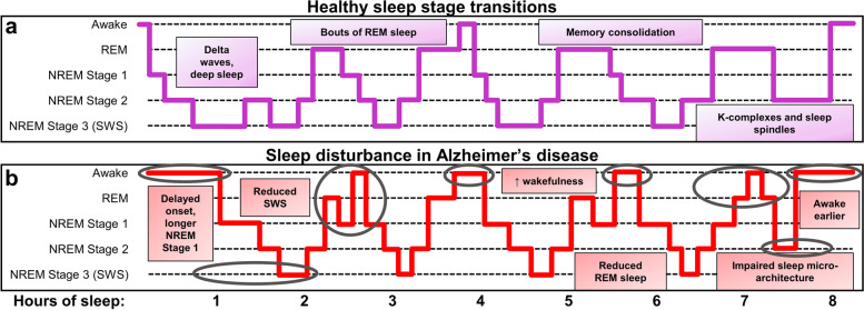
Failed proteostasis is a well-documented feature of Alzheimer's disease, particularly, reduced protein degradation and clearance. However, the contribution of failed proteostasis to neuronal circuit dysfunction is an emerging concept in neurodegenerative research and will prove critical in understanding cognitive decline. Our objective is to convey Alzheimer's disease progression with the growing evidence for a bidirectional relationship of sleep disruption and proteostasis failure. Proteostasis dysfunction and tauopathy in Alzheimer's disease disrupts neurons that regulate the sleep-wake cycle, which presents behavior as impaired slow wave and rapid eye movement sleep patterns. Subsequent sleep loss further impairs protein clearance. Sleep loss is a defined feature seen early in many neurodegenerative disorders and contributes to memory impairments in Alzheimer's disease. Canonical pathological hallmarks, β-amyloid, and tau, directly disrupt sleep, and neurodegeneration of locus coeruleus, hippocampal and hypothalamic neurons from tau proteinopathy causes disruption of the neuronal circuitry of sleep. Acting in a positive-feedback-loop, sleep loss and circadian rhythm disruption then increase spread of β-amyloid and tau, through impairments of proteasome, autophagy, unfolded protein response and glymphatic clearance. This phenomenon extends beyond β-amyloid and tau, with interactions of sleep impairment with the homeostasis of TDP-43, α-synuclein, FUS, and huntingtin proteins, implicating sleep loss as an important consideration in an array of neurodegenerative diseases and in cases of mixed neuropathology. Critically, the dynamics of this interaction in the neurodegenerative environment are not fully elucidated and are deserving of further discussion and research. Finally, we propose sleep-enhancing therapeutics as potential interventions for promoting healthy proteostasis, including β-amyloid and tau clearance, mechanistically linking these processes. With further clinical and preclinical research, we propose this dynamic interaction as a diagnostic and therapeutic framework, informing precise single- and combinatorial-treatments for Alzheimer's disease and other brain disorders.

Keywords: Alzheimer’s disease; Autophagy; Proteostasis; Sleep; Tau; Unfolded protein response; β-amyloid.


Conflict of interest statement

The authors declare that they have no competing interests.

Figures

Fig. 1
Fig. 1
Schematic of sleep disturbances in Alzheimer’s disease. Sleep is subdivided into stages of rapid eye movement (REM) and non-REM (NREM) sleep by signatures of neuronal activity. NREM can be further subdivided into 3 stages; NREM stage 3 is often referred to as slow wave sleep (SWS). a In healthy individuals, sleep begins in NREM stage 1, with waning neuronal activity and frequency, which further slows in restorative NREM stage 2 and SWS. SWS dominates early in the sleep cycle with synchronous, low frequency delta waves, whereas transitions to REM sleep occur a few hours after sleep onset, in ~ 90-min cycles. REM sleep electroencephalogram (EEG) is more akin to wakefulness with higher frequency and lower amplitude signals than SWS and dominated by theta waves. Memory consolidation is facilitated by bouts of REM, as well as NREM stage 2, prominent late in the sleep cycle, with characteristic high amplitude K-complexes and high frequency sleep spindles in EEG. In summary, REM and NREM stage 2 and 3 are important in memory consolidation [–68]; whereas SWS is also critical for toxic protein clearance and to reduce net synaptic strength to dampen aberrant plasticity and preserve a healthy signal:noise ratio of neuronal activity [68, 69]. Individuals who experience sleep disturbances are at a higher risk for Alzheimer’s disease (AD), and, moreover, those with AD exhibit characteristic features of sleep loss. b In AD, sleep is disrupted throughout the night, in which there is a delayed onset, longer bouts of non-restorative NREM stage 1 sleep, reduced bouts of SWS, REM and NREM stage 2, as well as increased wakefulness (notable changes compared to healthy sleep are circled). In sum, sleep disturbance poses a significant risk for AD and other neurodegenerative diseases, most prominently through dysregulation of mechanisms that facilitate proteinopathy and cognitive deficits (see Fig. 3). Panels A and B are schematic representations of healthy sleep and common disturbances that occur in AD. Healthy control sleep stages were informed from [68], and the results of the meta-analysis in [22] informed the AD impairments demonstrated in panel B
Fig. 2
Fig. 2
Summary of the sleep–wake circuitry and impact on NREM, REM and wake states. Briefly, neuromodulation from cholinergic (REM-active, wake-active), noradrenergic (wake and arousal) and serotonergic (in general wake-promoting, neuromodulatory sleep-promoting functions) neurons signals to the hypothalamus and ascending pathways to regulate the sleep–wake balance. Hypothalamic orexinergic and histaminergic neurons promote wake, and MCH promotes sleep. GABAergic (VLPO, POA, PZ) and glutamatergic (PB, BF, PPT/LDT) neurons facilitate sleep- and wake-states, respectively; though GABA can be wake-promoting in certain instances. See Sect. "Sleep-regulating centers and neurodegeneration" for further details [–172]. Regions are not to scale nor laid out anatomically. Arrows indicate activation signal to the efferent region and flat ends indicate inhibitory signal. Synaptic connections are colored by behavioral state: black dashed lines (ascending neuromodulatory activity with broad effects), red (wake and/or arousal), light red (wake- and REM-active), and blue (NREM and/or SWS). Abbreviations: acetylcholine (ACh); basal forebrain (BF); dorsal raphe nucleus (DRN); glutamate (glut); histamine (hist); lateral hypothalamus (LH); median preoptic nucleus (MnPO); melanin-concentrating hormone (MCH); noradrenaline (NA); non-rapid eye movement sleep (NREM); parabrachial nucleus (PB); parafacial zone (PZ); parvalbumin (PVB); pedunculopontine and laterodorsal tegmental nuclei (PPT/LDT); preoptic area (POA); polysomnography (PSG); rapid eye movement (REM); serotonin (5-HT); slow wave sleep (SWS) somatostatin (SST); suprachiasmatic nucleus (SCN); tuberomammillary nucleus (TMN); vasoactive intestinal polypeptide (VIP); vasopressin (VP); ventrolateral preoptic area (VLPO). Created with BioRender.com
Fig. 3
Fig. 3
Proteostasis of Aβ and tau is disrupted by Alzheimer’s-related sleep loss, driving proteinopathy, neuronal network dysfunction and cognitive impairment. Sleep is intimately linked to homeostatic processes that control protein accumulation, and when disturbed, can exacerbate and trigger proteinopathy. a Shows decrease in SWS and a concomitant decline in the metabolite clearance which is usually highest during SWS. Glymphatics involve brain influx of cerebrospinal fluid (CSF), travelling by bulk flow along periarterial spaces, which crosses the blood–brain barrier (BBB) via an astrocytic AQP4-mediated process, mixes with brain interstitial fluid (ISF), metabolites and solutes, and is cleared along perivenous spaces, driven by vasomotive forces. Bulk CSF/ISF efflux along veins drives metabolite clearance to dural lymphatic systems. Acutely, loss of SWS impairs glymphatic-mediated clearance of β-amyloid (Aβ) and tau, which chronically can feedback in cerebral amyloid angiopathy (CAA), tortuosity, enlarged perivascular spaces, and reduced blood flow, furthering glymphatic disruptions and increasing extracellular protein levels [226, 227]. b Sleep disturbance, circadian arrhythmicity, age and AD pathology all impact cellular proteostasis, contributing to an in general overactivation to clear protein; however, in cases of disease, proteostasis is overwhelmed and this activation exacerbates an already damaged system. BiP, and active levels of PERK, IRE1 and ATF6 are increased with sleep loss indicating UPR recruitment, which is insufficient to clear misfolded protein in aged- and diseased-states (indicated by red dashed line). Autophagy activation via Beclin-1 and atg4a, leads to nucleation and upregulated formation of autophagosomes (grey vacuoles), yet with a failure of autophagic flux there is reduced lysosomal (red vacuoles) fusion (indicated by red line). Notably, this can reduce Aβ and tau degradation, impart neurodegeneration through abundant axonal and dendritic autophagosomes, and promote proteinopathy through exosomal release of autophagosomes, as is seen in Alzheimer’s disease (AD) progression. Autophagy is regulated on a circadian cycle, and further impaired when this rhythm is disturbed. UPS failure occurs with disease state contributing to higher levels of intracellular protein that the ALP is unable to compensate for (indicated by red dashed line to p62). Dysregulated UPS-mediated degradation (indicated by red dashed line) of PERIOD proteins (including PER1 and PER2) may further circadian alterations. c During periods of prolonged wakefulness, higher frequency neuronal activity without restorative sleep promotes Aβ and tau cell-to-cell spread. Because of elevated synaptic strength, the neuronal signal:noise ratio decreases and synaptic plasticity saturates, leading to non-specific network activity [69]. Without rest, these potentially aberrant neuronal connections, in consort with accumulation of extra- and intracellular uncleared protein, exacerbate neuronal dysfunction, and cognitive processes such as memory can become impaired. d Finally, memory consolidation is impaired from loss of REM and NREM stage 2 and 3 sleep, contributing to transient memory loss. Neuronal activity of NREM UP- (i.e., spindles, sharp-wave ripples) and DOWN- (i.e., delta waves, K-complexes) states and REM theta oscillations consolidate memory circuits formed throughout the day [–68]. Chronically, impairments in proteostasis can progress to rampant accumulation of Aβ and tau in plaques and tangles, respectively, increasing disease spread and neuronal network dysfunction, all of which can further impair sleep and drive cognitive decline. Red text indicates impairments/decreases in AD and sleep disruption, green text indicates increases with AD and sleep disruption. Created with BioRender.com
Fig. 4
Fig. 4
Neuronal control of the sleep–wake cycle and therapeutic targets for sleep restoration in Alzheimer’s disease. The sleep–wake cycle is controlled by neuronal populations vulnerable in Alzheimer’s disease (AD). Circadian rhythmicity is mediated by hypothalamic neuronal activity and melatonin release from the pineal gland, normally maintaining a healthy sleep–wake cycle. Sleep is promoted by activity of melanin-concentrating hormone (MCH)-neurons in the hypothalamus and broad (including cortical, hippocampal, hypothalamic) GABAergic inhibitory signals. In the sleep-state, protein clearance and memory consolidation, mediated by entorhinal-hippocampal circuitry, are enhanced. Conversely, wakefulness and arousal are promoted by activity of histaminergic and orexinergic neurons of the hypothalamus, and noradrenergic neurons in the locus coeruleus. In the wake-state, cognitive and memory processes (mediated by entorhinal-hippocampal circuitry) occur with higher rates of neuronal activity, which potentiates Aβ and tau spread. Therapeutics to enhance sleep in AD present a unique opportunity to simultaneously improve the behavioral phenotype and reduce proteinopathy by improved proteostatic clearance. Enhancement of GABA signalling with pharmacological and non-pharmacological interventions may broadly improve network dysfunction in AD, for memory and sleep circuits. Notably, gamma entrainment is a novel and non-invasive strategy. Sleep promotion and balancing of circadian arrhythmicity can be accomplished via supplementation of the biologically active hormone melatonin, or non-pharmacological lifestyle interventions, including behavioral, light, music, and other auditory therapies. Pharmacological antihistamines and orexin antagonists decrease wake/arousal-signals and promote sleep. Potential exists for targeting of additional neuronal pathways to promote sleep, including noradrenergic signaling which is affected early in AD; the α2 adrenergic receptor agonist dexmedetomidine has been tested (see Table 2), but is more suitable as a sedative than therapeutic. Furthermore, the antidepressant trazodone has potential for improving sleep in AD acting through neuromodulation of serotonergic, adrenergic, histaminergic, and cholinergic pathways, as well as modifying the UPR. Future work is necessary to characterize and discover new sleep- and proteostasis-targeted therapies in AD. See Tables 2 and 3 for sleep-related AD clinical trials and their relevance to proteostasis. Created with BioRender.com

References

    1. Boland B, Yu WH, Corti O, Mollereau B, Henriques A, Bezard E, et al. Promoting the clearance of neurotoxic proteins in neurodegenerative disorders of ageing. Nat Rev Drug Discov. 2018;17:660–688. doi: 10.1038/nrd.2018.109. - DOI - PMC - PubMed
    1. Selkoe DJ, Hardy J. The amyloid hypothesis of Alzheimer’s disease at 25 years. EMBO Mol Med. 2016;8:595–608. doi: 10.15252/emmm.201606210. - DOI - PMC - PubMed
    1. Braak H, Braak E. Neuropathological stageing of Alzheimer-related changes. Acta Neuropathol. 1991;82:239–259. doi: 10.1007/BF00308809. - DOI - PubMed
    1. Hanseeuw BJ, Betensky RA, Jacobs HIL, Schultz AP, Sepulcre J, Becker JA, et al. Association of Amyloid and Tau With Cognition in Preclinical Alzheimer Disease: A Longitudinal Study. JAMA Neurol. 2019;76:915. doi: 10.1001/jamaneurol.2019.1424. - DOI - PMC - PubMed
    1. Busche MA, Wegmann S, Dujardin S, Commins C, Schiantarelli J, Klickstein N, et al. Tau impairs neural circuits, dominating amyloid-β effects, in Alzheimer models in vivo. Nat Neurosci. 2019;22:57–64. doi: 10.1038/s41593-018-0289-8. - DOI - PMC - PubMed

Publication types