

Polyglucosan body disease in an aged chimpanzee (Pan troglodytes)
- PMID: 37086019
- PMCID: PMC10642523
- DOI: 10.1111/neup.12906
Polyglucosan body disease in an aged chimpanzee (Pan troglodytes)
Abstract
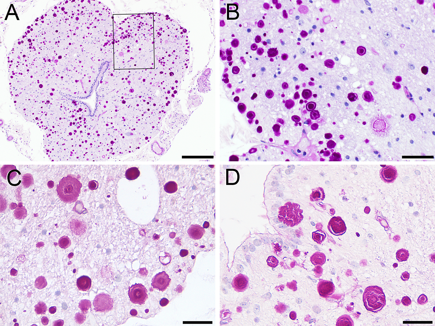
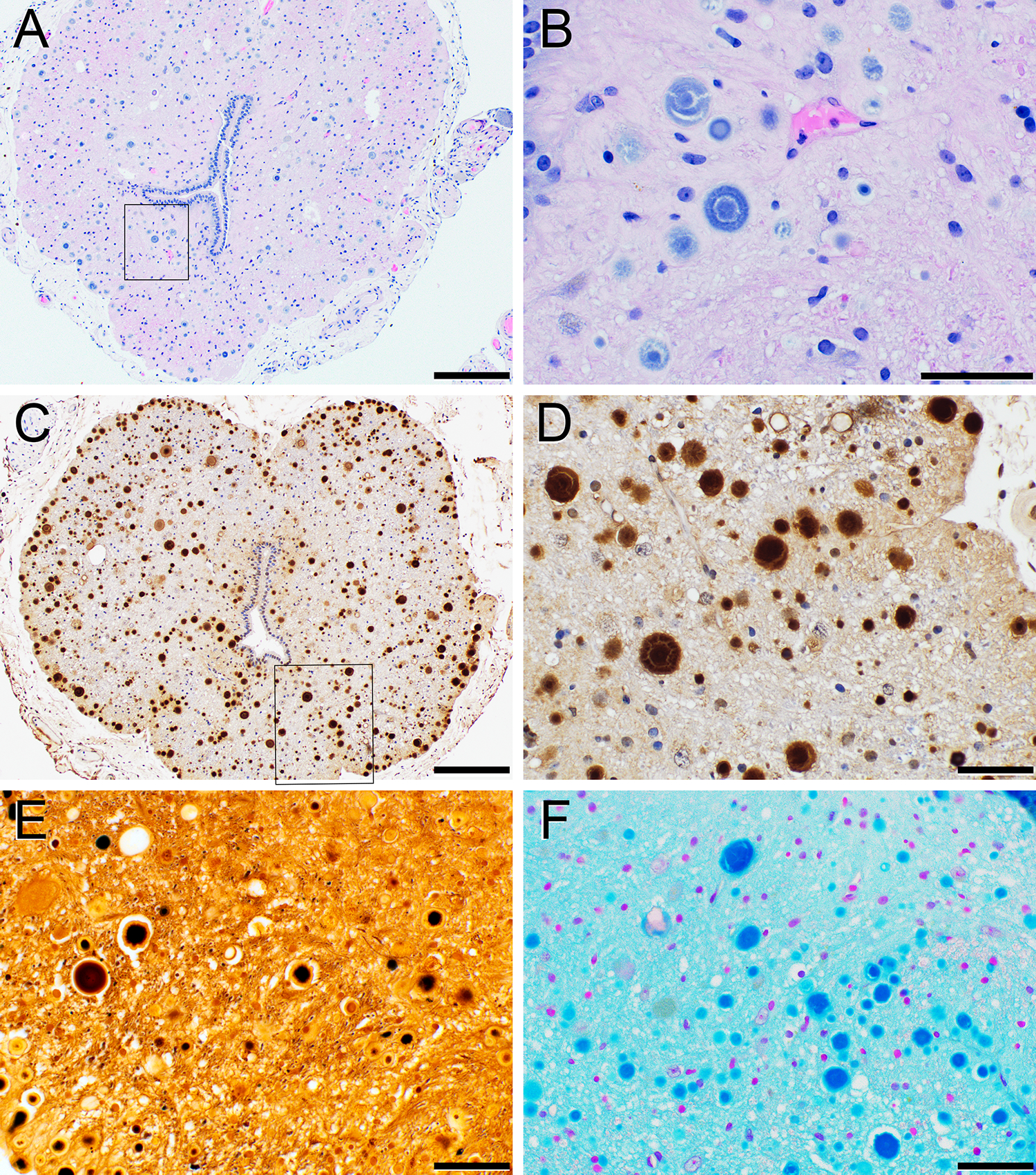
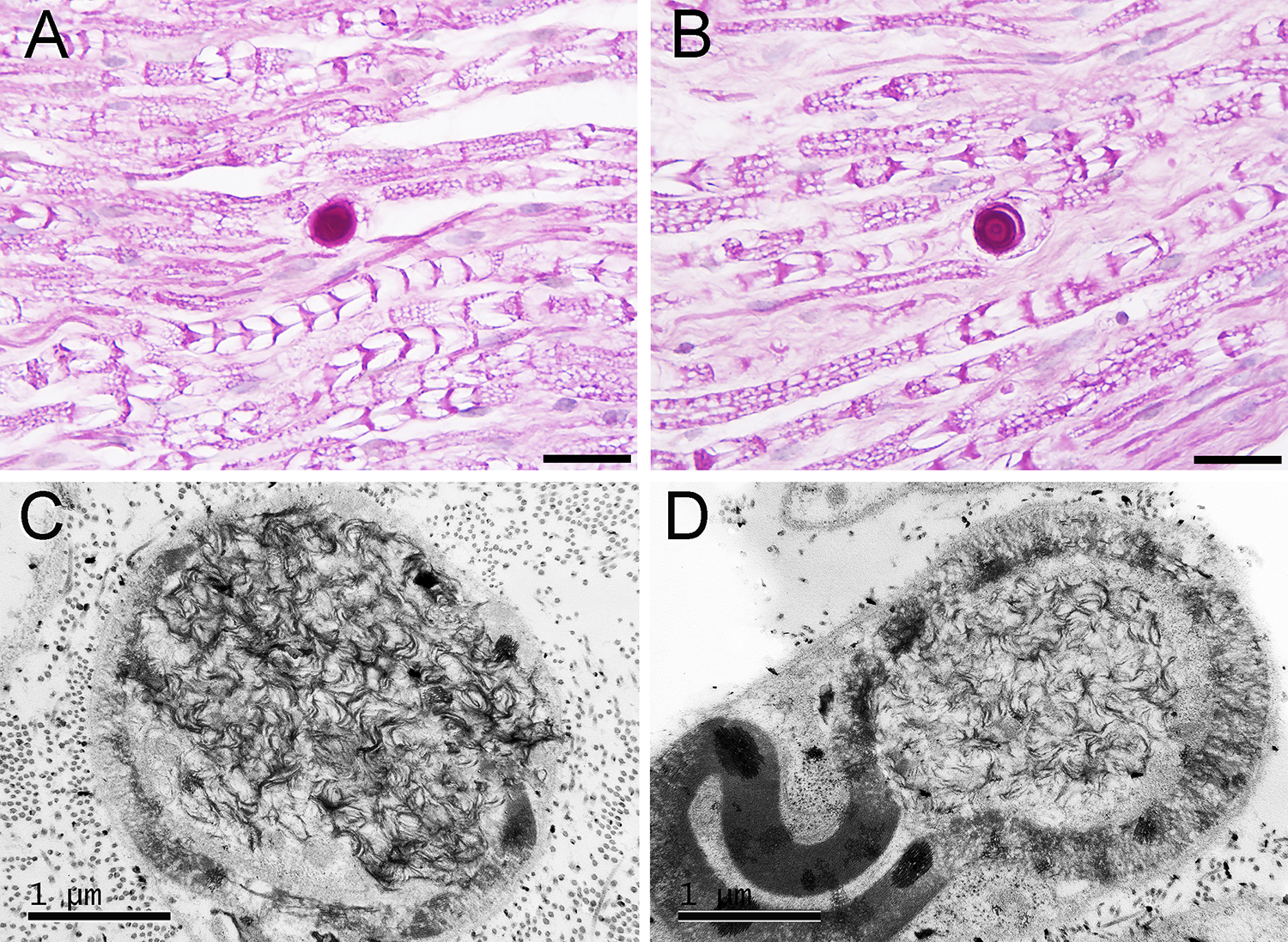
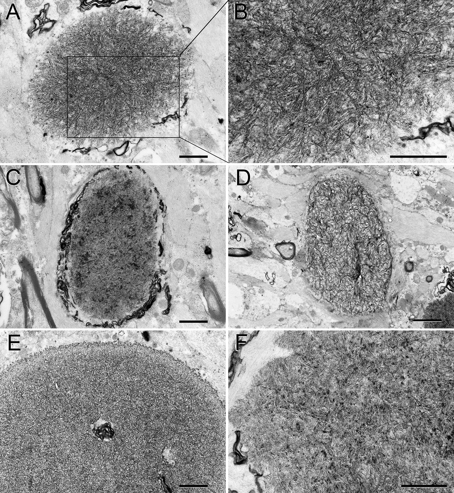
A 57-year-old female chimpanzee presented with a brief history of increasing lethargy and rapidly progressive lower-limb weakness that culminated in loss of use. Postmortem examination revealed no significant gross lesions in the nervous system or other organ systems. Histological analysis revealed round, basophilic to amphophilic polyglucosan bodies (PGBs) in the white and gray matter of the cervical, thoracic, lumbar, and coccygeal regions of spinal cord. Only rare PGBs were observed in forebrain samples. The lesions in the spinal cord were polymorphic, and they were positively stained with hematoxylin, periodic acid Schiff, Alcian blue, toluidine blue, Bielschowsky silver, and Grocott-Gomori methenamine-silver methods, and they were negative for von Kossa and Congo Red stains. Immunohistochemical evaluation revealed reactivity with antibodies to ubiquitin, but they were negative for glial fibrillary acidic protein, neuron-specific enolase, neurofilaments, tau protein, and Aβ protein. Electron microscopy revealed non-membrane-bound deposits composed of densely packed filaments within axons and in the extracellular space. Intra-axonal PGBs were associated with disruption of the axonal fine structure and disintegration of the surrounding myelin sheath. These findings are the first description of PGBs linked to neurological dysfunction in a chimpanzee. Clinicopathologically, the disorder resembled adult PGB disease in humans.
Keywords: Lafora bodies; adult polyglucosan body disease (APBD); aging; corpora amylacea; neurodegenerative disease.
© 2023 Japanese Society of Neuropathology.
Conflict of interest statement
DISCLOSURE
The authors declare no conflicts of interest for this report.
Figures
Similar articles
-
Characterisation of Lafora-like bodies and other polyglucosan bodies in two aged dogs with neurological disease.Vet J. 2010 Feb;183(2):222-5. doi: 10.1016/j.tvjl.2008.10.002. Epub 2008 Nov 14. Vet J. 2010. PMID: 19010069
-
Polyglucosan bodies in the brain of a cow.Acta Neuropathol. 1994;88(1):75-7. doi: 10.1007/BF00294362. Acta Neuropathol. 1994. PMID: 7941976
-
Histological, immunohistochemical and ultrastructural features of polyglucosan bodies in uterine smooth muscle of pet rabbits (Oryctolaguscuniculus).J Comp Pathol. 2023 Feb;201:28-32. doi: 10.1016/j.jcpa.2022.12.009. Epub 2023 Jan 18. J Comp Pathol. 2023. PMID: 36669389
-
Adult polyglucosan body myopathy with subclinical peripheral neuropathy: case report and review of diseases associated with polyglucosan body accumulation.Clin Neuropathol. 1988 Nov-Dec;7(6):271-9. Clin Neuropathol. 1988. PMID: 2852083 Review.
-
Corpora-amylacea and the family of polyglucosan diseases.Brain Res Brain Res Rev. 1999 Apr;29(2-3):265-95. doi: 10.1016/s0165-0173(99)00003-x. Brain Res Brain Res Rev. 1999. PMID: 10209236 Review.
References
-
- Cavanagh JB. Corpora-amylacea and the family of polyglucosan diseases. Brain Res Brain Res Rev. 1999;29(2–3):265–95. - PubMed
-
- Manich G, Cabezon I, Auge E, Pelegri C, Vilaplana J. Periodic acid-Schiff granules in the brain of aged mice: From amyloid aggregates to degenerative structures containing neo-epitopes. Ageing Res Rev. 2016;27:42–55. - PubMed
-
- Sakai M, Austin J, Witmer F, Trueb L. Studies of corpora amylacea. I. Isolation and preliminary characterization by chemical and histochemical techniques. Arch Neurol. 1969;21(5):526–44. - PubMed
-
- Seilhean D, De Girolami U, Gray F. Basic pathology of tne central nervous system. In: Gray F, Duyckaerts C, De Girolami U, editors. Escourolle and Poirier’s Manual of Basic Neuropathology. Oxford: Oxford University Press; 2014. p. 1–19.
Publication types
MeSH terms
Substances
Grants and funding
LinkOut - more resources
Full Text Sources
