. 2023 Jul;50(9):2621-2635.
doi: 10.1007/s00259-023-06211-6. Epub 2023 Apr 22.

Fibroblast activation protein targeted radiotherapy induces an immunogenic tumor microenvironment and enhances the efficacy of PD-1 immune checkpoint inhibition

Affiliations

Fibroblast activation protein targeted radiotherapy induces an immunogenic tumor microenvironment and enhances the efficacy of PD-1 immune checkpoint inhibition

Dirk Zboralski et al. Eur J Nucl Med Mol Imaging. 2023 Jul.

Abstract

Purpose: FAP is a membrane-bound protease under investigation as a pan-cancer target, given its high levels in tumors but limited expression in normal tissues. FAP-2286 is a radiopharmaceutical in clinical development for solid tumors that consists of two functional elements: a FAP-targeting peptide and a chelator used to attach radioisotopes. Preclinically, we evaluated the immune modulation and anti-tumor efficacy of FAP-2287, a murine surrogate for FAP-2286, conjugated to the radionuclide lutetium-177 (177Lu) as a monotherapy and in combination with a PD-1 targeting antibody.

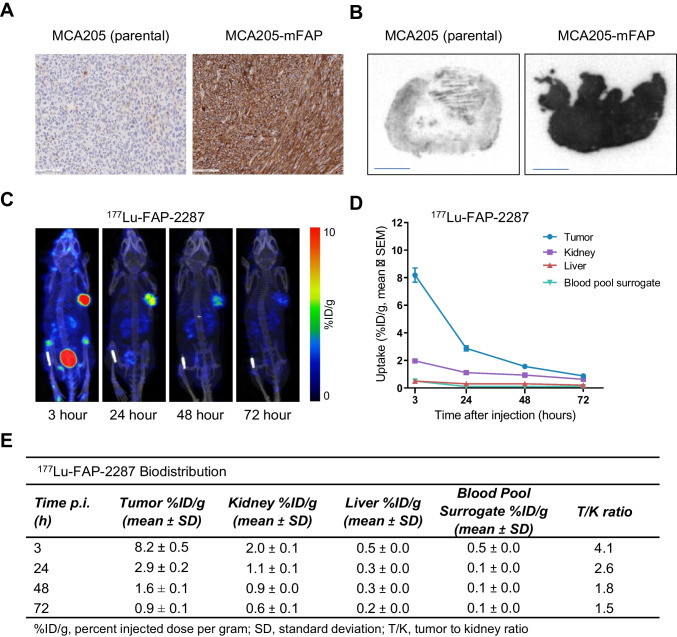
Methods: C57BL/6 mice bearing MCA205 mouse FAP-expressing tumors (MCA205-mFAP) were treated with 177Lu-FAP-2287, anti-PD-1, or both. Tumor uptake of 177Lu- FAP-2287 was assessed by SPECT/CT scanning, while therapeutic efficacy was measured by tumor volume and survival. Immune profiling of tumor infiltrates was evaluated through flow cytometry, RNA expression, and immunohistochemistry analyses.

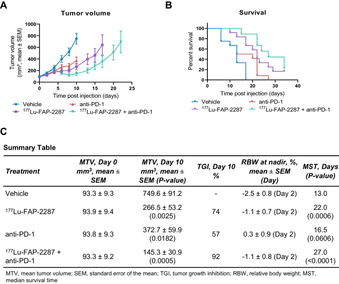
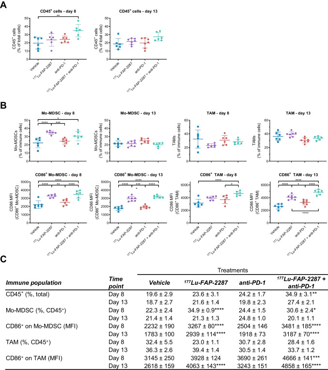
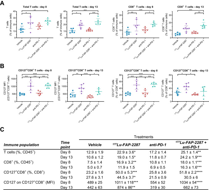
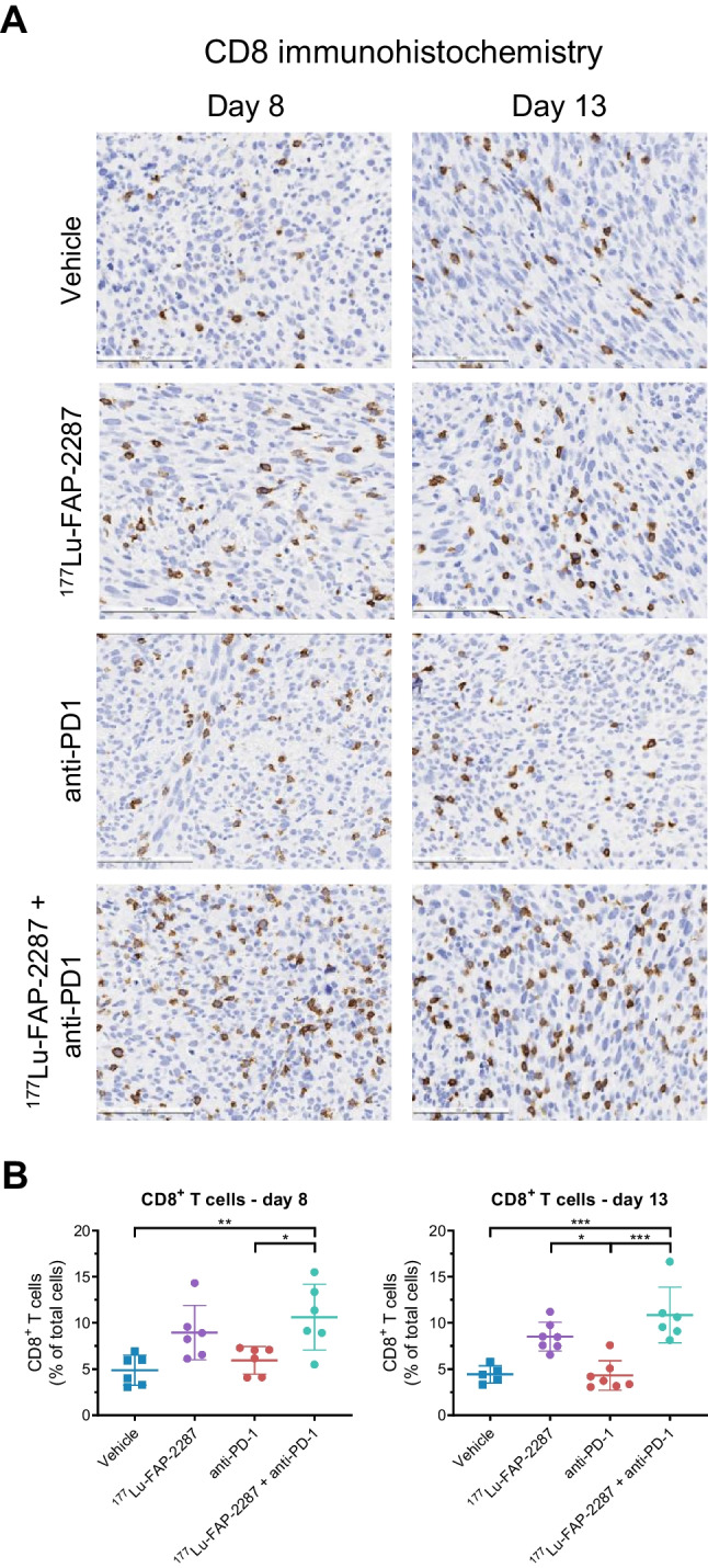
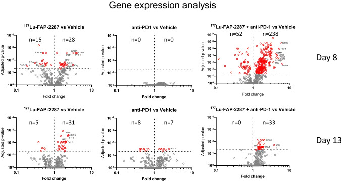
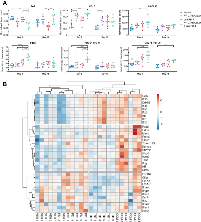
Results: 177Lu-FAP-2287 rapidly accumulated in MCA205-mFAP tumors leading to significant tumor growth inhibition (TGI) and longer survival time. Significant TGI was also observed from anti-PD-1 and the combination. In flow cytometry analysis of tumors, 177Lu-FAP-2287 increased CD8+ T cell infiltration which was maintained in the combination with anti-PD-1. The increase in CD8+ T cells was accompanied by an induction of STING-mediated type I interferon response and higher levels of co-stimulatory molecules such as CD86.

Conclusion: In a preclinical model, FAP-targeted radiotherapy enhanced anti-PD-1-mediated TGI by modulating the TME and increasing the recruitment of tumor-infiltrating CD8+ T cells. These findings provide a rationale for clinical studies of combined 177Lu-FAP-2286 radiotherapy and immune checkpoint inhibition in FAP-positive tumors.

Keywords: CD8; FAP; PD-1; STING; TRT; Theranostic.


Conflict of interest statement

D. Zboralski, F. Osterkamp, A. Bredenbeck, A. Schumann, A. Hoehne, E. Schneider, M. Paschke, J. Ungewiss, C. Haase are employees of 3B Pharmaceuticals GmbH and are named inventors of FAP-2287. M. Paschke, and F. Osterkamp are cofounders of 3B Pharmaceuticals GmbH. E. Christensen is an employee of Minerva Imaging ApS. L. Robillard, A.D. Simmons, T.C. Harding and M. Nguyen are employees of Clovis Oncology, Inc., and may own stock or have stock options in the company.

Figures

Fig. 1
Fig. 1
In vivo uptake of 177Lu-FAP-2287 into FAP-positive MCA205-mFAP syngeneic tumors. Representative images of MCA205-mFAP tumors analyzed by FAP IHC (A, 200 μm) and 111In-FAP-2287 ARG (B, 2 mm) are shown. SPECT images of one representative mouse at 4 different timepoints are shown (C). Quantification of 177Lu-FAP-2287 biodistribution as mean ± SEM %ID/g (n = 6) in tumor and kidney at 3, 24, 48 and 72 h pi (D) and summary table of uptake in several organs and tumor to kidney ratios (E) are shown
Fig. 2
Fig. 2
Tumor efficacy of 177Lu-FAP-2287 and in combination with anti-PD-1 in MCA205-mFAP syngeneic tumor model. MCA205-mFAP tumor-bearing mice were treated with vehicle, 177Lu-FAP-2287, anti-PD-1 antibody, or combination 177Lu-FAP-2287 plus anti-PD-1 (n = 12 mice/group). Mean tumor volumes ± SEM during study period (A), Kaplan Meier survival curve (B) and a summary table (C) are shown
Fig. 3
Fig. 3
Immune profiling of CD86+ myeloid subsets after treatment with 177Lu-FAP-2287 and in combination with anti-PD-1 in MCA205-mFAP syngeneic tumor model. Individual percentage of CD45+ cells with bars as mean ± SEM on day 8 and 13 pi (A), individual percentage of Mo-MDSC and TAM and their CD86 MFI with bars as mean ± SEM on day 8 and 13 pi (B), and a summary table (C) are shown. Significant changes compared to vehicle in the summary table and between groups in the graph are denoted as * p < 0.05, ** p < 0.01, *** p < 0.001 and **** p < 0.0001
Fig. 4
Fig. 4
Immune profiling of CD8+ T cells after treatment with 177Lu-FAP-2287 and in combination with anti-PD-1 in MCA205-mFAP syngeneic tumor model. Individual percentage of T cells and CD8+ T cells with bars as mean ± SEM on day 8 and 13 pi (A), individual percentage of CD127+CD8+ cells and their CD127 MFI with bars as mean ± SEM on day 8 and 13 pi (B), and a summary table (C) are shown. Significant changes compared to vehicle in the summary table and between groups in the graph are denoted as * p < 0.05, ** p < 0.01, *** p < 0.001 and **** p < 0.0001
Fig. 5
Fig. 5
IHC analysis of CD8+ T cells after treatment with 177Lu-FAP-2287 and in combination with anti-PD-1 in MCA205-mFAP syngeneic tumor model. Representative images show CD8 staining in MCA205-mFAP tumors (A, 100 μm) and individual percentage of CD8+ cells by IHC quantification with bars as mean ± SEM are plotted for days 8 and 13 pi (B). Significant changes between groups are denoted as * p < 0.05, ** p < 0.01, *** p < 0.001 and **** p < 0.0001
Fig. 6
Fig. 6
Differential expression analysis of MCA205-mFAP tumors after treatment with 177Lu-FAP-2287 and in combination with anti-PD-1. Fold changes in gene expression versus vehicle are shown as volcano plots with statistically significant genes above the horizontal line, and highly differentially expressed genes on either side
Fig. 7
Fig. 7
Molecular profiling of MCA205-mFAP tumors after treatment with 177Lu-FAP-2287 and in combination with anti-PD-1. Individual normalized linear count with bars as mean ± SEM are plotted for FAP and several immune genes on days 8 and 13 pi (A). Significant changes between groups are denoted as * p < 0.05, ** p < 0.01, *** p < 0.001 and **** p < 0.0001. The heatmap was obtained by hierarchical clustering centered around genes with correlation distance and complete linkage, using ClustVis tools (https://bio.tools/clustvis). The color scale displays the row z-score: red color indicates high abundance while blue color low abundance (B). Groups are marked as VEH, vehicle; TRT, 177Lu-FAP-2287; PD1, anti-PD-1, COM, combination

References

    1. Darvin P, Toor SM, Sasidharan Nair V, Elkord E. Immune checkpoint inhibitors: recent progress and potential biomarkers. Exp Mol Med. 2018;50:1–11. doi: 10.1038/s12276-018-0191-1. - DOI - PMC - PubMed
    1. Schoenfeld AJ, Hellmann MD. Acquired Resistance to Immune Checkpoint Inhibitors. Cancer Cell. 2020;37:443–455. doi: 10.1016/j.ccell.2020.03.017. - DOI - PMC - PubMed
    1. Sharma P, Hu-Lieskovan S, Wargo JA, Ribas A. Primary, Adaptive, and Acquired Resistance to Cancer Immunotherapy. Cell. 2017;168:707–723. doi: 10.1016/j.cell.2017.01.017. - DOI - PMC - PubMed
    1. Joyce JA, Fearon DT. T cell exclusion, immune privilege, and the tumor microenvironment. Science. 2015;348:74–80. doi: 10.1126/science.aaa6204. - DOI - PubMed
    1. Sahai E, Astsaturov I, Cukierman E, DeNardo DG, Egeblad M, Evans RM, et al. A framework for advancing our understanding of cancer-associated fibroblasts. Nat Rev. 2020;20:174–186. doi: 10.1038/s41568-019-0238-1. - DOI - PMC - PubMed

Substances