. 2023 Jun:259:106539.
doi: 10.1016/j.aquatox.2023.106539. Epub 2023 Apr 12.

Exposure to the persistent organic pollutant 2,3,7,8-tetrachlorodibenzo-p-dioxin (TCDD, dioxin) disrupts development of the zebrafish inner ear

Affiliations

Exposure to the persistent organic pollutant 2,3,7,8-tetrachlorodibenzo-p-dioxin (TCDD, dioxin) disrupts development of the zebrafish inner ear

Layra G Cintrón-Rivera et al. Aquat Toxicol. 2023 Jun.

Abstract

Dioxins are a class of highly toxic and persistent environmental pollutants that have been shown through epidemiological and laboratory-based studies to act as developmental teratogens. 2,3,7,8-tetrachlorodibenzo-p-dioxin (TCDD), the most potent dioxin congener, has a high affinity for the aryl hydrocarbon receptor (AHR), a ligand activated transcription factor. TCDD-induced AHR activation during development impairs nervous system, cardiac, and craniofacial development. Despite the robust phenotypes previously reported, the characterization of developmental malformations and our understanding of the molecular targets mediating TCDD-induced developmental toxicity remains limited. In zebrafish, TCDD-induced craniofacial malformations are produced, in part, by the downregulation of SRY-box transcription factor 9b (sox9b), a member of the SoxE gene family. sox9b, along with fellow SoxE gene family members sox9a and sox10, have important functions in the development of the otic placode, the otic vesicle, and, ultimately, the inner ear. Given that sox9b is a known target of TCDD and that transcriptional interactions exist among SoxE genes, we asked whether TCDD exposure impaired the development of the zebrafish auditory system, specifically the otic vesicle, which gives rise to the sensory components of the inner ear. Using immunohistochemistry, in vivo confocal imaging, and time-lapse microscopy, we assessed the impact of TCDD exposure on zebrafish otic vesicle development. We found exposure resulted in structural deficits, including incomplete pillar fusion and altered pillar topography, leading to defective semicircular canal development. The observed structural deficits were accompanied by reduced collagen type II expression in the ear. Together, our findings reveal the otic vesicle as a novel target of TCDD-induced toxicity, suggest that the function of multiple SoxE genes may be affected by TCDD exposure, and provide insight into how environmental contaminants contribute to congenital malformations.

Keywords: Dioxin; Ear; Otic vesicle; Semicircular canal; TCDD; Zebrafish.


Conflict of interest statement

Declaration of Competing Interest The authors declare no conflicts of interest. This study was completed without any financial affiliations that could be interpreted as a potential conflict of interest.

Figures

Fig. 1.
Fig. 1.. Anatomy of the wild-type zebrafish ear.
Schematics of wild-type zebrafish ear at 48 hpf (A) and 72 hpf (E). Representative brightfield images of live wild-type ears showing the developing cristae and otoliths at 48 hpf (B-D). Representative brightfield images of live wild-type ears capturing the cristae, otoliths, pillars, and dorsolateral septum at 72 hpf (F-H). Abbreviations: anterior crista (AC), ventral crista (VC), posterior crista (PC), anterior pillar (AP), ventral pillar (VP), posterior pillar (PP), dorsolateral septum (DLS), anterior otolith (AO), posterior otolith (PO). All images are 40x. Scale bar in B & F is 25μm.
Fig. 2.
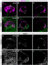
Fig. 2.. TCDD exposure expands the Cyp1a expression domain in the developing embryonic ear at 48 hpf.
Confocal micrographs of control (A-C) and TCDD-exposed (D-F) embryos carrying reporters for sox10 (magenta) and cyp1a (green) [Tg(sox10:RFP); TgBAC(cyp1a:NLS:EGFP)]. sox10 expression encompasses the otic epithelium and cyp1a acts as a biomarker of AHR activation. Representative slices taken from confocal z-series capturing cyp1a induction (white) in control (G-I) and TCDD-exposed (J-L) embryos. Maximum intensity projections of confocal z-series are shown in A, D, G, & J. Abbreviations: anterior crista (AC), ventral crista (VC), and posterior crista (PC). Scale bar in A, D, G, & J = 25μm. Total n in the control group=8 and n in the TCDD-exposed group=10. Embryos and larvae were collected across 3 replicates.
Fig. 3.
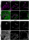
Fig. 3.. TCDD-induced expansion of Cyp1a expression is maintained in the larval ear at 72 hpf.
Confocal micrographs of control (A-C) and TCDD-exposed (D-F) larvae carrying reporters for sox10 (magenta) and cyp1a (green) [Tg(sox10:RFP); Tg(cyp1a:NLS:EGFP)]. Cyp1a transgenic reporter in black and white in controls (G-I) and TCDD exposed (J-L) fish. Maximum intensity projections of confocal z-series are shown in A, D, G, & J. Abbreviations: anterior crista (AC), ventral crista (VC), posterior crista (PC), anterior pillar (AP), ventral pillar (VP), posterior pillar (PP), dorsolateral septum (DLS). Scale bar in A, D, G & J is 25μm. Total n in the control group=10 and n in the TCDD-exposed group=13. Embryos and larvae were collected across 3 replicates.
Fig. 4.
Fig. 4.. Quantification of structural deficits induced by TCDD exposure.
Distribution of coincident absent structures in control and TCDD exposed larvae at 72 hpf (A). Percentage of specific ear structure presence versus absence (B). Abbreviations: anterior pillar (AP), ventral pillar (VP), posterior pillar (PP), dorsolateral septum (DLS). Total n in the control group=20 and n in the TCDD-exposed group=24. Embryos and larvae were collected across 3 replicates. ****p < 0.0001.
Fig. 5.
Fig. 5.. Quantification of incomplete pillar fusion.
Percent of structures that failed to establish a connection at fusion plate in control and TCDD exposed larvae at 72hpf (A). Abbreviations: anterior pillar (AP), ventral pillar (VP), posterior pillar (PP). Total n in the control group=20 and n in the TCDD-exposed group=24. Embryos and larvae were collected across 3 replicates. ****p < 0.0001.
Fig. 6.
Fig. 6.. Pillar topography defect quantification and phenotypes.
Distribution of control and TCDD exposed larvae with zero, one, or two displaced pillars at 72 hpf (A). Confocal slices capturing the fluorescent reporter Tg(sox10:RFP), in magenta, in the developing otic epithelium at 72hpf with a 40x magnification (B-G). Control ear displaying typical pillar topography (B-D). TCDD exposed ear exhibiting displacement of both the anterior and posterior pillar (E-G). Abbreviations: anterior pillar (AP), ventral pillar (VP), posterior pillar (PP), dorsolateral septum (DLS), anterior crista (AC), posterior crista (PC). 25μm scale bar for reference (B,E). Total n in the control group=20 and n in the TCDD-exposed group=24. Embryos and larvae were collected across 3 replicates.
Fig. 7.
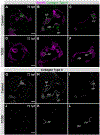
Fig. 7.. Loss of collagen type II expression in the ear following TCDD exposure.
40x confocal micrographs slices highlighting different portions of the developing ear at 72 hpf (A-L). Control (A-C) and TCDD exposed larvae (D-F) expressing the fluorescent reporter Tg(sox10:RFP) (magenta) and immunostained for collagen type II (green). Black and white slices of collagen type II in control (G-I) and TCDD exposed (J-L) larval ears. Abbreviations: fusion plate (FP), anterior macula (AM), anterior pillar (AP), ventral pillar (VP), posterior pillar (PP). Scale bar in A,D,G,J = 25μm. Total n in the control group=11 and n in the TCDD-exposed group=8. Embryos and larvae were collected across 3 replicates.

Update of

References

    1. Abbas L, Whitfield TT, 2010. The zebrafish inner ear. Fish Physiol. 29 (C), 123–171. 10.1016/S1546-5098(10)02904-3. - DOI
    1. Acke FRE, Dhooge IJM, Malfait F, De Leenheer EMR, 2012. Hearing impairment in Stickler syndrome: a systematic review. Orphanet. J. Rare. Dis 7 (1), 1. 10.1186/1750-1172-7-84. - DOI - PMC - PubMed
    1. Andreasen EA, Mathew LK, Löhr CV, Hasson R, Tanguay RL, 2007. Aryl hydrocarbon receptor activation impairs extracellular matrix remodeling during zebra fish fin regeneration. Toxicol. Sci 95 (1), 215–226. 10.1093/toxsci/kfl119. - DOI - PubMed
    1. Andreasen EA, Mathew LK, Tanguay RL, 2006. Regenerative growth is impacted by TCDD: gene expression analysis reveals extracellular matrix modulation. Toxicol. Sci 92 (1), 254–269. 10.1093/toxsci/kfj118. - DOI - PubMed
    1. Barrionuevo F, Naumann A, Bagheri-Fam S, Speth V, Taketo MM, Scherer G, Neubüser A, 2008. Sox9 is required for invagination of the otic placode in mice. Dev. Biol 317 (1), 213–224. 10.1016/j.ydbio.2008.02.011. - DOI - PubMed

MeSH terms

LinkOut - more resources