. 2023 May 22;58(10):825-835.e6.
doi: 10.1016/j.devcel.2023.03.019. Epub 2023 Apr 21.

Extracellular matrix assembly stress initiates Drosophila central nervous system morphogenesis

Affiliations

Extracellular matrix assembly stress initiates Drosophila central nervous system morphogenesis

Eduardo Serna-Morales et al. Dev Cell. .

Abstract

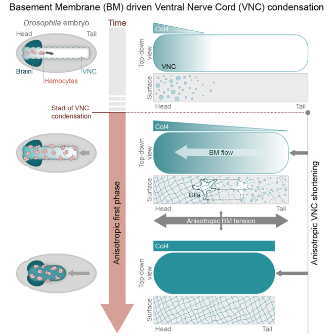
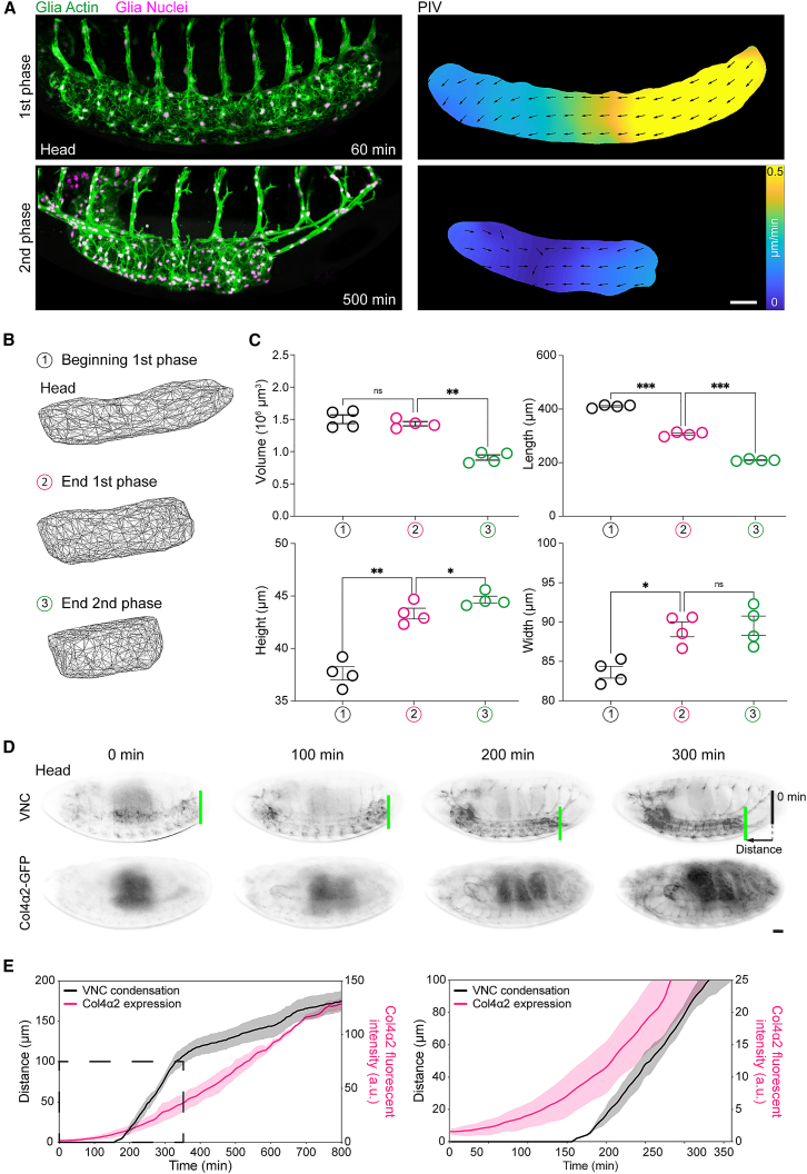
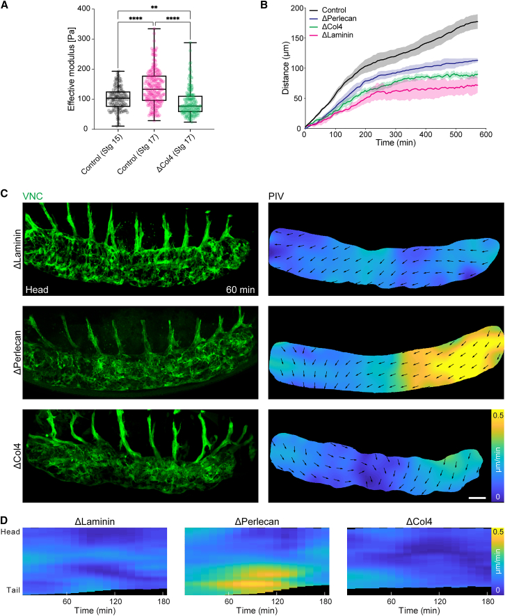
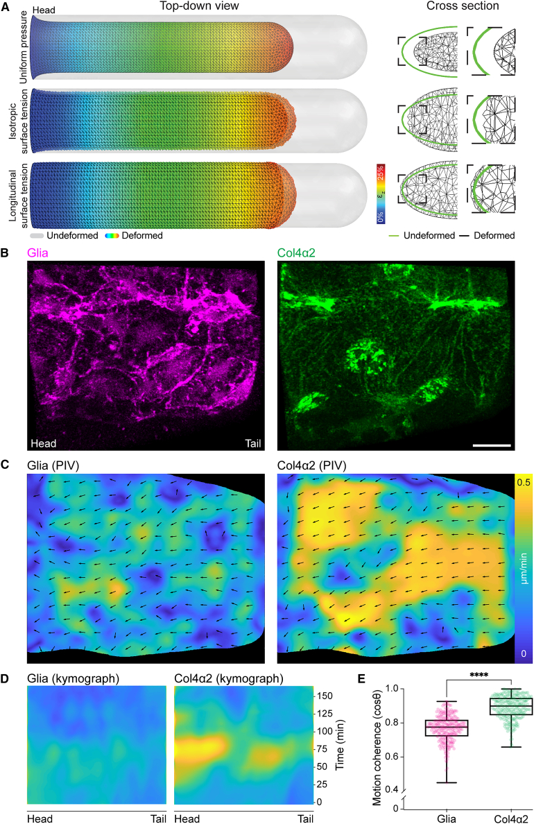
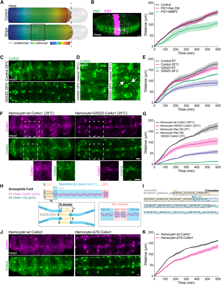
Forces controlling tissue morphogenesis are attributed to cellular-driven activities, and any role for extracellular matrix (ECM) is assumed to be passive. However, all polymer networks, including ECM, can develop autonomous stresses during their assembly. Here, we examine the morphogenetic function of an ECM before reaching homeostatic equilibrium by analyzing de novo ECM assembly during Drosophila ventral nerve cord (VNC) condensation. Asymmetric VNC shortening and a rapid decrease in surface area correlate with the exponential assembly of collagen IV (Col4) surrounding the tissue. Concomitantly, a transient developmentally induced Col4 gradient leads to coherent long-range flow of ECM, which equilibrates the Col4 network. Finite element analysis and perturbation of Col4 network formation through the generation of dominant Col4 mutations that affect assembly reveal that VNC morphodynamics is partially driven by a sudden increase in ECM-driven surface tension. These data suggest that ECM assembly stress and associated network instabilities can actively participate in tissue morphogenesis.

Keywords: Drosophila; basement membrane; central nervous system; collagen IV; embryonic development; extracellular matix; morphogenesis; surface tension.


Conflict of interest statement

Declaration of interests The authors declare no competing interests.

Figures

None
Graphical abstract
Figure 1
Figure 1
VNC morphogenesis involves distinct stages and correlates with the initiation of Col4 assembly (A) Live imaging of VNC morphogenesis (left panels) and quantification of tissue motion by PIV (right panels) revealing anisotropic (1st phase) and isotropic (2nd phase) phases of condensation. Scale bar, 30 μm. (B) 3D reconstruction of VNC shape during the phases of condensation. (C) Quantification of VNC shape at the start (1), end of the 1st phase (2) and end of the 2nd phase (3) of condensation. Repeated measures one-way ANOVA with Geisser-Greenhouse correction and Holm-Šídák’s multiple comparisons tests. Each dot represents one embryo, n= 4 embryos. Error bars show standard error of the mean. Volume: ns p = 0.4130, ∗∗p = 0.0073; Length: ∗∗∗p = 0.0004; Height: ∗∗p = 0.0045, ∗p = 0.0155; Width: ∗p = 0.0278, ns p = 0.6803. (D) Live imaging of VNC condensation (highlighted by the difference between the green and black lines) and induction of Col4 production by quantifying fluorescence intensity of Col4α2-GFP. Scale bar, 30 μm. (E) Correlation of Col4 fluorescence intensity with the rate of VNC condensation as measured by tracking the motion of the tail of the tissue. Right panel focuses on the data within the dashed square. n = 3 embryos. See also Video S1.
Figure 2
Figure 2
Col4 induction correlates with an increase in VNC stiffness, and the loss of Col4 inhibits the anisotropic phase of VNC condensation (A) Atomic force microscopy of the VNC revealing an increase in stiffness from stage 15 to 17 (1st and 2nd phases, respectively), which is lost in the absence of Col4. Kruskal-Wallis tests and Dunn’s multiple comparisons tests. n = 137 indentations (one control embryo, stg 15), n = 207 (two control embryos, stg 17), n = 181 (two ΔCol4 embryos, stg 17). Each dot represents one indentation. Boxplots show medians, 25th and 75th percentiles as box limits, 10th and 90th percentiles as whiskers. ∗∗∗∗p < 0.0001, ∗∗p = 0.0018. (B) Quantification of VNC condensation rate by tracking tail motion as in Figures 1D and 1E in control (n = 4 embryos) and laminin, perlecan, and col4 mutant embryos (n = 3 embryos each). (C) Live imaging of the 1st phase of VNC condensation as in Figure 1A in laminin, perlecan, and col4 mutants. Scale bar, 30 μm. (D) Kymograph of the average speed of VNC condensation from PIV analysis in (C) highlighting an absence of an anisotropic phase of condensation in laminin and col4 mutants. See also Video S2.
Figure 3
Figure 3
Anisotropy in surface tension and coherent long-range flow of Col4 is sufficient to explain the sudden isovolumetric change in VNC shape (A) Finite element analysis (FEA) of VNC morphogenesis. Simulations of VNC deformation assuming (top panel) uniform normal surface pressure, (middle panel) uniform increase in surface tension, or (bottom panel) anisotropic increase in surface tension along the length of the tissue. Only anisotropic surface tension leads to reduction in length and increase in height and width of the tissue. (B) Live imaging of glia and Col4 motion on the VNC surface. Scale bar, 10 μm. (C) Simultaneous tracking of glia and Col4 motion by PIV. (D) Kymograph of the PIV analysis in (C) highlighting a sudden increase in Col4 speed during VNC condensation which is not observed by tracking glia. (E) Correlation of local alignment of PIV vectors reveals that motion of the Col4 network is more coherent than glial motion. Mann-Whitney test. n = 256 vectors each. Each dot represents one PIV vector. Boxplots show medians, 25th and 75th percentiles as box limits, 10th and 90th percentiles as whiskers. ∗∗∗∗p < 0.0001. See also Video S3.
Figure 4
Figure 4
Direct perturbations of Col4 assembly slow the rate of VNC condensation (A) FEA of VNC condensation in which surface tension is locally reduced in a stripe in the middle of the tissue. Simulations suggest that local perturbation of surface tension leads to long-range reduction in deformation of the tail of the tissue. (B) Local disruption of the ECM network by MMP2 expression in a central parasegment (PS7, n = 4), in contrast to control (n = 3) and expression of Rac DN (n = 3), is sufficient to affect the rate of VNC condensation as measured by tracking the tail of the tissue as in Figures 1D and 1E. Scale bar, 30 μm. (C) Live imaging of Col4α2-GFP reveals that rearing embryos containing a temperature-sensitive (TS) point mutation in Col4α1 (G552D) at the non-permissive temperature (29°C) inhibits BM network assembly. Scale bar, 10 μm. (D) Rearing the TS mutant at the permissive temperature (18°C) to allow some ECM assembly and switching to the non-permissive temperature (29°C) leads to aggregation of extracellular Col4 (arrows) and accumulation of soluble Col4 in the hemocoel (asterisks) showing that the TS mutant affects the Col4 network. Scale bar, 10 μm. (E) Quantification of VNC condensation in the TS mutant reveals that the condensation rate can be affected by temperature titration (RT, room temperature). Control data reused from Figure 2B. n = 4 control RT and 29°C, n = 5 G552D RT and 29°C. (F) Expression of wild-type or G552D point mutant Col4α1 in hemocytes during VNC condensation in the background of endogenously GFP-tagged Col4α2. Bottom panels are high-magnification views of the highlighted regions revealing that the G552D transgene fails to incorporate into the ECM network and inhibits incorporation of Col4α2. Scale bars, 10 μm. (G) Quantification of VNC condensation in the genotypes highlighted in (F) reveals that hemocyte-specific expression of the G552D transgene slows VNC condensation. Furthermore, expression of the G552D mutant in hemocytes that also express Rac DN is sufficient to rescue the severe phenotype. n = 3 Hemocyte>wt Col4α1, Hemocyte>Rac DN; G552D Col4α1, n = 5 Hemocyte>G552D Col4α1; Hemocyte>Rac DN is from Figure S3C. (H) Schematic highlighting Col4 domains and interactions hypothesized to drive network assembly. In the C terminus of Col4 are interactions between NC1 domains, which lead to dimerization of Col4 trimers. At the N terminus are interactions of a putative 7S domain, which consists of the first non-helical (NH1) and triple-helical (TH1) regions of the protein. Disulfide bonds and noncovalent interactions between 7S domains are hypothesized to lead to tetramerization of Col4 trimers. Sp, signal peptide. (I) Amino acid sequence of the N terminus of Drosophila Col4. The dashed line highlights the amino acid sequence truncated from the putative 7S domain of Col4α1 to generate a dominant negative transgene. (J) Expression of wild-type (WT) Col4α1 or a truncation of the 7S domain in hemocytes shows that deletion of the 7S domain decreases its incorporation and reduces incorporation of Col4α2. Scale bar, 10 μm. (K) Quantification of VNC condensation after expression of the transgenes in (H) reveals that removal of the 7S domain slows the rate of motion of the tail of the tissue. n = 4 embryos each. See also Video S2.

Comment in

References

    1. Gilbert S.F. Sixth Edition. Sinauer Associates; 2000. Developmental Biology.
    1. Walma D.A.C., Yamada K.M. The extracellular matrix in development. Development. 2020;147 doi: 10.1242/dev.175596. - DOI - PMC - PubMed
    1. Munjal A., Hannezo E., Tsai T.Y., Mitchison T.J., Megason S.G. Extracellular hyaluronate pressure shaped by cellular tethers drives tissue morphogenesis. Cell. 2021;184:6313–6325.e18. doi: 10.1016/j.cell.2021.11.025. - DOI - PMC - PubMed
    1. Loganathan R., Rongish B.J., Smith C.M., Filla M.B., Czirok A., Bénazéraf B., Little C.D. Extracellular matrix motion and early morphogenesis. Development. 2016;143:2056–2065. doi: 10.1242/dev.127886. - DOI - PMC - PubMed
    1. Newman S.A., Frenz D.A., Tomasek J.J., Rabuzzi D.D. Matrix-driven translocation of cells and nonliving particles. Science. 1985;228:885–889. doi: 10.1126/science.4001925. - DOI - PubMed

Publication types

LinkOut - more resources