. 2023 Jun;299(6):104736.
doi: 10.1016/j.jbc.2023.104736. Epub 2023 Apr 21.

Augmin is a Ran-regulated spindle assembly factor

Affiliations

Augmin is a Ran-regulated spindle assembly factor

Jodi Kraus et al. J Biol Chem. 2023 Jun.

Abstract

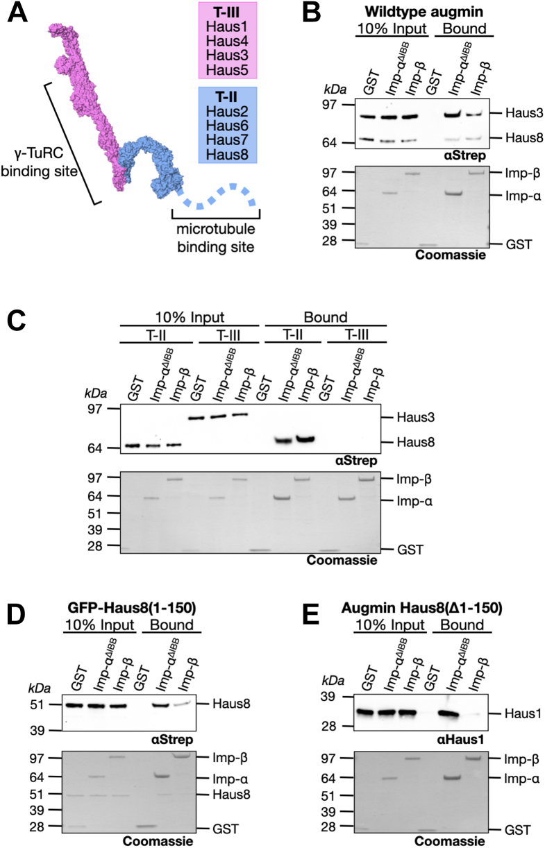
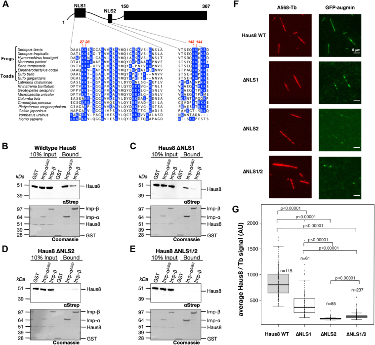
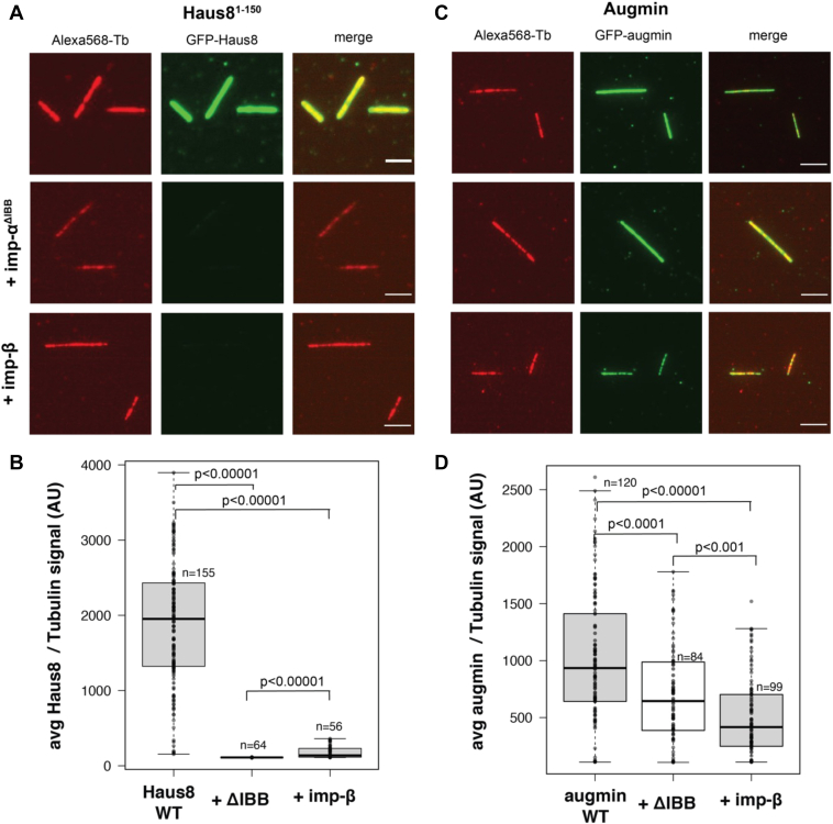
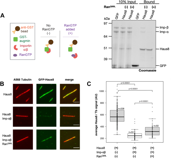
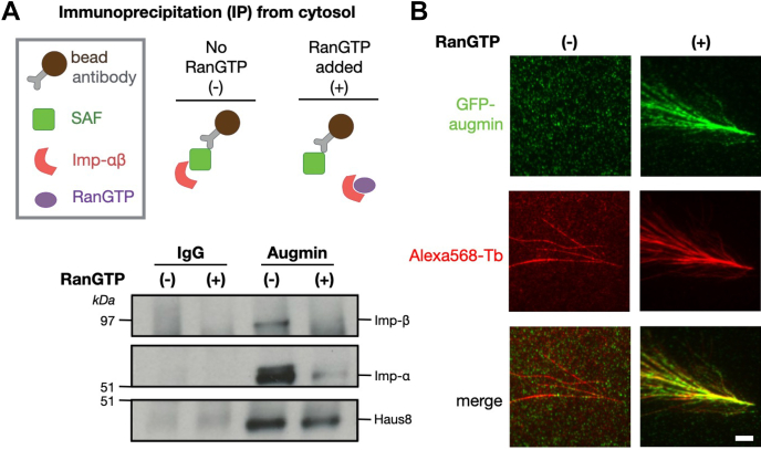
Mitotic spindles are composed of microtubules (MTs) that must nucleate at the right place and time. Ran regulates this process by directly controlling the release of spindle assembly factors (SAFs) from nucleocytoplasmic shuttle proteins importin-αβ and subsequently forms a biochemical gradient of SAFs localized around chromosomes. The majority of spindle MTs are generated by branching MT nucleation, which has been shown to require an eight-subunit protein complex known as augmin. In Xenopus laevis, Ran can control branching through a canonical SAF, TPX2, which is nonessential in Drosophila melanogaster embryos and HeLa cells. Thus, how Ran regulates branching MT nucleation when TPX2 is not required remains unknown. Here, we use in vitro pulldowns and total internal reflection fluorescence microscopy to show that augmin is a Ran-regulated SAF. We demonstrate that augmin directly interacts with both importin-α and importin-β through two nuclear localization sequences on the Haus8 subunit, which overlap with the MT-binding site. Moreover, we show that Ran controls localization of augmin to MTs in both Xenopus egg extract and in vitro. Our results demonstrate that RanGTP directly regulates augmin, which establishes a new way by which Ran controls branching MT nucleation and spindle assembly both in the absence and presence of TPX2.

Keywords: Ran pathway; TPX2; augmin; branching microtubule nucleation; importins; spindle assembly factor.


Conflict of interest statement

Conflict of interest The authors declare that they have no conflicts of interest with the contents of this article.

Figures

Figure 1
Figure 1
Augmin binds to importins.A, structure of the augmin complex, broken into the γ-TuRC binding T-III (pink), comprised of subunits Haus1 and Haus3–5, and the MT-binding T-II (blue), comprised of subunits Haus2 and Haus6–8. The disordered N terminus of Haus8 (shown as a dashed blue line) contains the primary MT-binding site. Structure of Xenopus augmin was taken from Ref. (39). B, glutathione beads bound to either GST (control), GST-importin-αΔIBB, or GST-importin-β were incubated with full-length augmin, then both the input and bound fraction were Western blotted for intact augmin complexes using an antibody against the Strep-tagged subunits Haus3 (T-III) and Haus8 (T-II). Below, GST and GST-importin loading was demonstrated by Coomassie stain. C, as in (B), importin-bound beads were incubated with augmin, either T-III or T-II, and binding of intact augmin subcomplex was detected via Western blot against the Strep-tagged subunits Haus3 and Haus8. D, Haus81–150 (fused to an N-terminal Strep-tagged GFP) was incubated with importin-bound beads, and binding was detected via Western blot. E, augmin complex lacking the N-terminal 150 residues of Haus8 was incubated with importin-bound beads, and binding of intact augmin complex was detected via Western blot against augmin subunit Haus1. γ-TuRC, γ-tubulin ring complex; GST, glutathione-S-transferase; IBB, importin-β binding; MT, microtubule; T-III, tetramer III.
Figure 2
Figure 2
Haus8 of augmin binds to importins and MTs through two NLS sites.A, the augmin subunit Haus8 is predicted to contain two NLS sequences within its disordered N terminus (43). The domain architecture of Haus8 is cartooned at top, and the sequence of each predicted NLS in Xenopus laevis is shown at the bottom. X. laevis Haus8 is shown aligned to other vertebrate orthologs below, and all basic residues (arginine abbreviated as R and lysine as K) are highlighted in blue. Indicated in red at the top of the sequence are the pairs of basic residues mutated to alanine to generate the Haus8 mutants ΔNLS1 (K27A/K28A) and ΔNLS2 (K143A/K144A). BE, pulldowns of Strep-GFP-Haus81–150, either wildtype or containing the indicated NLS mutants, were conducted as for Figure 1D. F, selected TIRF images of in vitro binding of GST-GFP-Haus81–150 to stabilized MT seeds. Haus8 constructs with mutated residues in NLS1 and/or NLS2 result in a reduction in binding, as quantified in G. Images belonging to the same experiment were contrast matched. To compare augmin fluorescence intensity across experiments, the intensity was normalized with respect to the tubulin signal. G, boxplot of average GFP-Haus8 signal relative to the average tubulin signal, where each marker represents a single MT from the experiment shown in F. The total number of MTs (n) was collected from two replicates. Center lines show the medians; box limits indicate the 25th and 75th percentiles as determined by R software; whiskers extend 1.5 times the interquartile range from the 25th and 75th percentiles. p Values were calculated from independent t tests. GST, glutathione-S-transferase; MT, microtubule; NLS, nuclear localization signal; TIRF, total internal reflection fluorescence.
Figure 3
Figure 3
Importins regulate augmin binding to MTs.A, WT GST-GFP-Haus8 localizes strongly to GMPCPP-stabilized MT seeds in vitro (top row), as visualized by TIRF microscopy. In the presence of importin-αΔIBB (middle row) or importin-β (bottom row), binding of Haus8 to MTs is diminished. This is quantified in B. B, boxplot of average GFP-Haus8 signal relative to the average tubulin signal, where each marker represents a single MT from the experiment shown in F. The total number of MTs (n) was collected from two replicates. The boxes extend from the 25th to 75th percentile, and the upper and lower bars represent the minimum and maximum, respectively. p Values were calculated from independent t tests. C, WT GFP-labeled augmin localized to GMPCPP-stabilized MT seeds in vitro (top row), as visualized by TIRF microscopy. In the presence of importin-αΔIBB (middle row) or importin-β (bottom row), binding of augmin to MTs is decreased but not eliminated. This is quantified in D. D, boxplot of average GFP-augmin signal relative to the average tubulin signal, where each marker represents a single MT from the experiment shown in F. The total number of MTs (n) was collected from two replicates using two independent augmin preparations. Center lines show the medians; box limits indicate the 25th and 75th percentiles as determined by R software; whiskers extend 1.5 times the interquartile range from the 25th and 75th percentiles. p Values were calculated from independent t tests. In A and C, images belonging to the same experiment were contrast matched. To compare Haus8 fluorescence intensity across experiments, the intensity was normalized with respect to the tubulin signal. Scale bars correspond to 5 μm. GST, glutathione-S-transferase; IBB, importin-β binding; MT, microtubule; TIRF, total internal reflection fluorescence.
Figure 4
Figure 4
RanGTP releases importin inhibition of MT binding.A, GFP or GFP-Haus81–150 was bound to α-GFP magnetic resin and incubated with importin-αβ in the presence or the absence of a 10-fold excess of RanQ69L. Both importin-αβ binding and augmin loading were assessed by Coomassie staining and the intensity of bands at the indicated sizes. B, in vitro localization of GST-GFP-Haus81–150 binding to GMPCPP-stabilized MT seeds, as visualized by TIRF microscopy (top row). Addition of importin-αβ heterodimer inhibits binding of Haus8 to MTs (middle row), whereas addition of RanQ69L rescues MT binding of Haus8 (bottom row). Images belonging to the same experiment were contrast matched. To compare Haus8 fluorescence intensity across experiments, the intensity was normalized with respect to the tubulin signal. This is quantified in C. C, boxplot of average GFP-Haus8 signal relative to the average tubulin signal, where each marker represents a single MT from the experiment shown in F. n corresponds to the total number of MTs. Center lines show the medians; box limits indicate the 25th and 75th percentiles as determined by R software; whiskers extend 1.5 times the interquartile range from the 25th and 75th percentiles. p Values were calculated from independent t tests. Scale bars correspond to 5 μm. GST, glutathione-S-transferase; MT, microtubule; TIRF, total internal reflection fluorescence.
Figure 5
Figure 5
Ran regulates augmin in Xenopus egg extract.A (top), schematic of immunoprecipitation (IP) strategy where antibodies were conjugated to magnetic beads (top). Bottom, Western blot of IPs for a control antibody (immunoglobulin [IgG]) and antiaugmin in the presence or the absence of RanQ69L. B, TIRF images of augmin in Xenopus egg extract in the absence (left column) and presence (right column) of RanQ69L. Branching MT nucleation reactions were carried out with Alexa568-labeled tubulin and fixed after 15 min. Endogenous augmin was detected via indirect immunofluorescence with custom primary antibodies against Haus8 and Alexa488-conjugated secondary antibodies. Scale bar corresponds to 5 μm. MT, microtubule; TIRF, total internal reflection fluorescence.

Similar articles

Cited by

References

    1. Meunier S., Vernos I. Acentrosomal microtubule assembly in mitosis: the where, when, and how. Trends Cell Biol. 2016;26:80–87. - PubMed
    1. Petry S. Mechanisms of mitotic spindle assembly. Annu. Rev. Biochem. 2016;85:659–683. - PMC - PubMed
    1. Wu J., Akhmanova A. Microtubule-organizing centers. Annu. Rev. Cell Dev. Biol. 2017;33:51–75. - PubMed
    1. Gunzelmann J., Ruthnick D., Lin T.C., Zhang W.L., Neuner A., Jakle U., et al. The microtubule polymerase Stu2 promotes oligomerization of the gamma-TuSC for cytoplasmic microtubule nucleation. Elife. 2018;7 - PMC - PubMed
    1. Oakley B.R., Paolillo V., Zheng Y.X. gamma-Tubulin complexes in microtubule nucleation and beyond. Mol. Biol. Cell. 2015;26:2957–2962. - PMC - PubMed

Publication types

MeSH terms

LinkOut - more resources