. 2023 Jun:179:90-101.
doi: 10.1016/j.yjmcc.2023.04.004. Epub 2023 Apr 20.

The role of P21-activated kinase (Pak1) in sinus node function

Affiliations

The role of P21-activated kinase (Pak1) in sinus node function

Carlos H Pereira et al. J Mol Cell Cardiol. 2023 Jun.

Abstract

Sinoatrial node (SAN) dysfunction (SND) and atrial arrhythmia frequently occur simultaneously with a hazard ratio of 4.2 for new onset atrial fibrillation (AF) in SND patients. In the atrial muscle attenuated activity of p21-activated kinase 1 (Pak1) increases the risk for AF by enhancing NADPH oxidase 2 dependent production of reactive oxygen species (ROS). However, the role of Pak1 dependent ROS regulation in SAN function has not yet been determined. We hypothesize that Pak1 activity maintains SAN activity by regulating the expression of the hyperpolarization activated cyclic nucleotide gated cation channel (HCN). To determine Pak1 dependent changes in heart rate (HR) regulation we quantified the intrinsic sinus rhythm in wild type (WT) and Pak1 deficient (Pak1-/-) mice of both sexes in vivo and in isolated Langendorff perfused hearts. Pak1-/- hearts displayed an attenuated HR in vivo after autonomic blockage and in isolated hearts. The contribution of the Ca2+ clock to pacemaker activity remained unchanged, but Ivabradine (3 μM), a blocker of HCN channels that are a membrane clock component, eliminated the differences in SAN activity between WT and Pak1-/- hearts. Reduced HCN4 expression was confirmed in Pak1-/- right atria. The reduced HCN activity in Pak1-/- could be rescued by class II HDAC inhibition (LMK235), ROS scavenging (TEMPOL) or attenuation of Extracellular Signal-Regulated Kinase (ERK) 1/2 activity (SCH772984). No sex specific differences in Pak1 dependent SAN regulation were determined. Our results establish Pak1 as a class II HDAC regulator and a potential therapeutic target to attenuate SAN bradycardia and AF susceptibility.

Keywords: Atrial fibrillation; HCN channel; Membrane clock; Reactive oxygen species; Sinoatrial node; p21-activated kinase 1.


Conflict of interest statement

Declaration of Competing Interests:

The authors have no competing interests.

Figures

Figure 1.
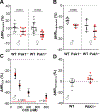
Figure 1.. Loss of Pak1 attenuates the intrinsic heart rate (HR) in male and female mice.
Representative recordings from male (●) and female (■) WT (n: ●=16, ■=16) and Pak1−/− (n: ●=13, ■=12) mice showing (Aa) ECGs in control conditions and (Ba) after suppression of autonomic signaling (atropine: 1 mg/kg + propranolol: 1 mg/kg), (n: ●=16, ■=13, ●=13, ■=8), (Ca) atrial electrograms from isolated hearts (n: ●=24, ■=14, ●=28, ■=12) as well as (Da) ECGs from isolated hearts paced at 8Hz (n: ●=4 ●=4), red arrows indicate the stimulation artifact. Quantification of the heart rate (HR) under the described conditions is shown in Ab - Cb, respectively. Quantification of the PR interval in isolated hearts is shown in Db. Data are presented as mean ± SD. One-Way ANOVA.
Figure 2.
Figure 2.. Alterations in ANS signaling mask the attenuated HR in Pak1−/− mice:
Quantification of the percentage change in HR in male (●) and female (■) WT (n: ●=10, ■=13) and Pak1−/− (n: ●=9, ■=8) mice in response to the suppression of (A) parasympathetic (atropine) and (B) sympathetic signaling (propranolol) (n: ●= 6, ●=4, ■= 5, ■=6). Percent change in HR of isolated male WT and Pak1−/− hearts in response to perfusion with (C) crescent CCh concentrations (sample size indicated in the figure for each CCh concentration) or (D) a blocker of GIRK, TerQ (n: ●=6 and ●=7). Data are means ± SD. Student’s t test (A, C - D) and Mann-Whitney test (B).
Figure 3:
Figure 3:. Loss of Pak1 attenuates the contribution of the voltage clock to pacemaker activity.
Representative HR recordings from isolated, Langendorff perfused male and female WT (●, ■) and Pak1−/− (●, ■) hearts during (A) superfusion with the SERCA inhibitor CPA or (B) the HCN4 inhibitor IVA. Summary data of CPA and IVA induced percent change in HR, respectively (Ab, n: ●=6, ●= 5, ■=3, ■= 4) and (Bb, n: ●=6, ●=6, ■=5, ■=6) as well as absolute HR in CPA (Ac, n: ●=6, ●=5, ■=3, ■=4) or IVA (Bc, n: ●=6, ●=6, ■=5, ■=6). Representative Western blot displaying HCN4 protein levels (Ca) and quantification of HCN4 protein levels normalized to actin expression (Cb). Data are means ± SD. One-Way ANOVA (A –B) Student’s t test (C).
Figure 4:
Figure 4:. Pak1 controls HCN contribution to pacemaker activity through class II HDACs.
HR analysis in male and female WT (●, ■) and Pak1−/− (●, ■) Langendorff perfused hearts under control conditions and after treatment of mice with LMK (WT: ◯, ◻; Pak1−/−: ◯,). Quantification of the basal HR (♂: Aa, n: ●= 24, ◯=5, ● =28, ◯=5) (♀: Ba, n: ◻=14, ◻=5, ■=12, ☐=4) and the percentage change in HR after perfusion with IVA (♂: Ab, n: ●=6, ◯=5, ● =6, ◯=4) (♀: Bb, n: =5, =4, =6, =4) or CPA (♂: Ac, n: ●=6, ◯=5, ● =5, ◯=4) and (♀: Bc, n: =3, =3, =4, =4). Data are presented as means ± SD. One Way ANOVA.
Figure 5.
Figure 5.. ROS-dependent regulation of HCN contribution to pacemaker activity.
HR analysis in WT (●) and Pak1−/− (●) Langendorff perfused hearts under control conditions and after treatment of mice with TEMPOL (WT: ◯; Pak1−/−: ). Quantification of the basal HR (A, n: ●= 38, ◯=7, ● =40, ◯=7) and percentage change in HR after perfusion with IVA (B, n: ●= 11, ◯=6, ● =12, ◯=7) or CPA (C, n: ●= 9, ◯=6, ● =9, ◯=4). (D) Change of cellular DCF fluorescence over time in atrial myocytes isolated from WT (●, n of cells/mice = 7/3, Pak1−/− (●, n = 10/3) or Pak1−/− hearts from mice treated with LMK2335 (◯, n = 7/2). Data are presented as means ± SD. One Way ANOVA (A –C), Nested ANOVA (D). * represents WT vs Pak1−/−, where p<0.05 *; # represents Pak1−/− vs Pak1−/− LMK, where p<0.05 #; p<0.01##.
Figure 6:
Figure 6:. Attenuation of Pak1 promotes increased activity of ERK and p38:
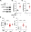
Representative Western blots displaying p-ERK1/2 and p-p38 levels in protein lysate from HL-1 cells after (Aa, n = 7/group) adenoviral gene transfer of LacZ or Pak1-siRNA. P-ERK1/2 (Ab) and p-p38 (Ac) protein levels normalized to GAPDH expression. Summary of experimental results from Langendorff perfused WT (●) and Pak1−/− (●) hearts under control conditions or after treatment with SCH772984 (ERKi; ◯, ◯). HR after ERKi treatment (B, n: ●=38, ◯=4, ● =40, ◯=5) and the IVA induced change in HR (C, n: ● =11 ◯=3 ● =12, ◯=5). Data are presented as mean ± SD. Student’s t test (A-B) and One Way ANOVA (C-D).
Figure 7.
Figure 7.. ROS but not SAN bradycardia promotes atrial arrhythmia in Pak1−/− hearts.
A: Representative traces of atrial electrograms showing the recovery of sinus rhythm (SR) after burst pacing (BP) in WT (black) and Pak1−/− (red) hearts. B: Percentage of WT and Pak1−/− animals with AF (◻, ☐) or SR (■, ■) after BP. C: Percentage of BP episodes eliciting AF/heart during control conditions (●=19, ●=13) and after LMK235 (◯=10, ◯=9), TEMPOL (◯=7, ◯=7) or SCH772984 (ERKi) (◯=4, ◯=5) treatment. Data are presented as mean ± SD. One Way ANOVA (C)

Similar articles

Cited by

References

    1. Choudhury M, Boyett MR, Morris GM, Biology of the Sinus Node and its Disease, Arrhythmia Electrophysiol Rev. 4 (2015) 28–34. 10.15420/aer.2015.4.1.28. - DOI - PMC - PubMed
    1. Egom EE, Vella K, Hua R, Jansen HJ, Moghtadaei M, Polina I, Bogachev O, Hurnik R, Mackasey M, Rafferty S, Ray G, Rose RA, Impaired sinoatrial node function and increased susceptibility to atrial fibrillation in mice lacking natriuretic peptide receptor C., J. Physiol. (Lond.). 593 (2015) 1127–1146. 10.1113/jphysiol.2014.283135. - DOI - PMC - PubMed
    1. Yang P-S, Kim D, Jang E, Yu HT, Kim T-H, Sung J-H, Pak H-N, Lee M-H, Joung B, Risk of sick sinus syndrome in patients diagnosed with atrial fibrillation: A population-based cohort., J Cardiovasc Electr. 32 (2021) 2704–2714. 10.1111/jce.15202. - DOI - PubMed
    1. DiFrancesco D, HCN4, Sinus Bradycardia and Atrial Fibrillation., Arrhythmia Electrophysiol Rev. 4 (2015) 9–13. 10.15420/aer.2015.4.1.9. - DOI - PMC - PubMed
    1. Elvan A, Wylie K, Zipes DP, Pacing-induced chronic atrial fibrillation impairs sinus node function in dogs. Electrophysiological remodeling., Circulation. 94 (1996) 2953–60. 10.1161/01.cir.94.11.2953. - DOI - PubMed

Publication types

Substances