. 2023 Apr 22;23(1):370.
doi: 10.1186/s12885-023-10858-7.

Prognostic significance of lymphovascular invasion in patients with pT1b esophageal squamous cell carcinoma

Affiliations

Prognostic significance of lymphovascular invasion in patients with pT1b esophageal squamous cell carcinoma

Linxiu Liu et al. BMC Cancer. .

Abstract

Background: Lymphovascular invasion (LVI) is a crucial predictor of lymph node metastasis (LNM). However, few studies have investigated the LVI positivity rate and its clinical significance in pT1b esophageal squamous cell carcinoma (ESCC) using immunohistochemistry and elastin staining.

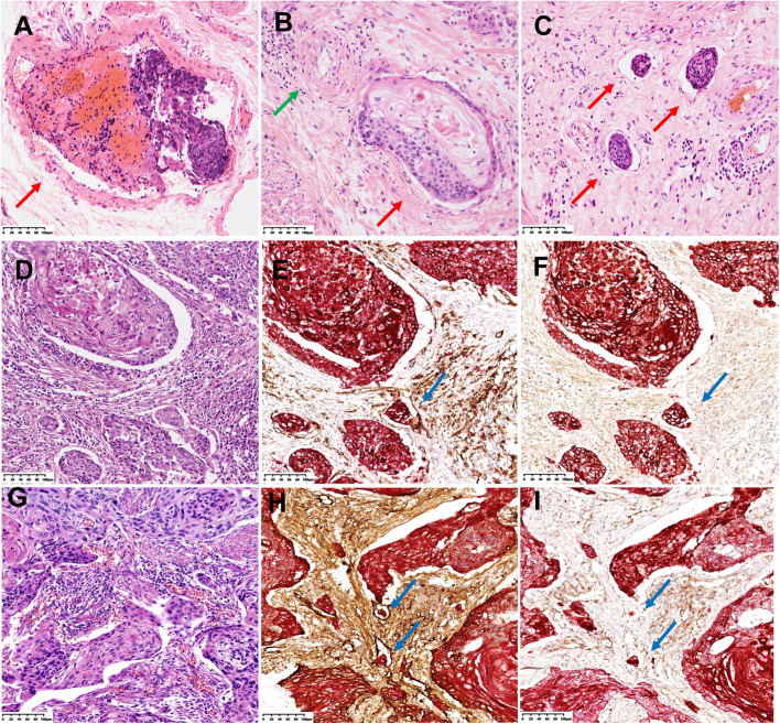
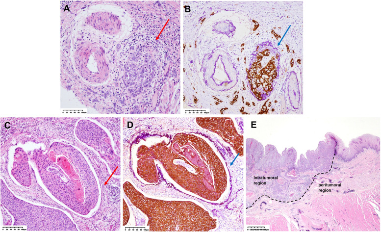
Methods: We collected data from158 patients with pT1b ESCC who had undergone radical esophagectomy. All paraffin blocks of invasive carcinoma from each patient were subjected to HE staining, elastin staining + CK (AE1/AE3) immunohistochemistry (E&IHC), and CD31/D2-40 + CK (AE1/AE3) double immunohistochemistry (D-IHC). The LVI was classified into types, i.e., vascular invasion (VI) and lymphatic vessel invasion (LI), and its location, quantity, and clinical significance were explored.

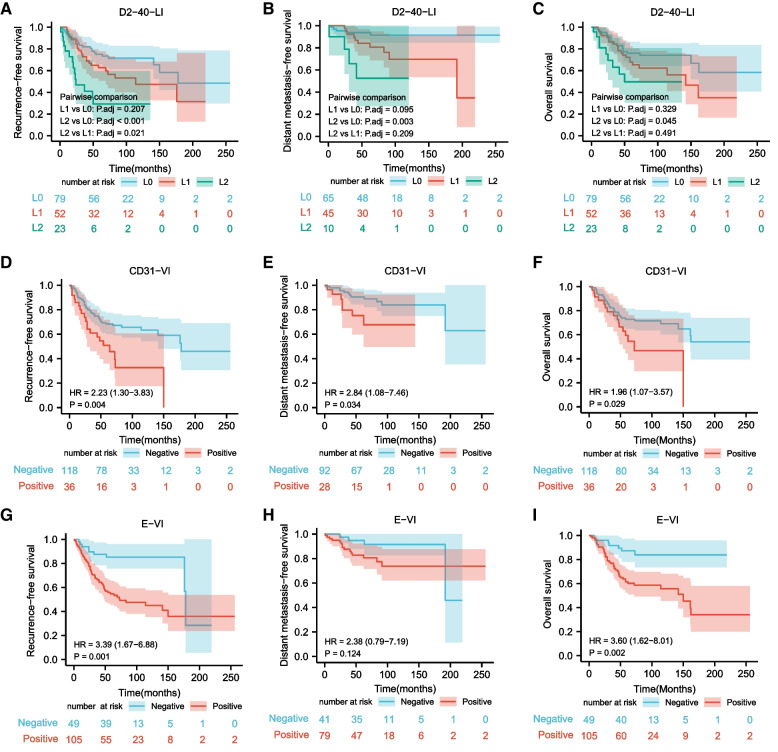
Results: The positivity rates of VI by E&IHC (E-VI), VI by CD31D-IHC (CD31-VI), and LI by D2-40 D-IHC (D2-40-LI) were significantly higher than those obtained by HE staining (P < 0.001, respectively). CD31-VI and E-VI were independent adverse prognostic factors for recurrence-free survival (RFS), and they were significantly associated with poor distant metastasis-free survival and overall survival in pT1b ESCC. Intratumoral LVI was also crucial in pT1b ESCC, and L2 (the count of D2-40-LI was 5 or more) was the strongest predictor for LNM and RFS in pT1b ESCC.

Conclusion: E&IHC and D-IHC can dramatically improve the detection rate of LVI in pT1b ESCC, and the classification and grading of LVI can help to improve the prediction of LNM and prognosis.

Keywords: Distant metastasis-free survival; Esophageal squamous cell carcinoma; Lymph node metastasis; Lymphovascular invasion; Overall survival; Recurrence-free survival.


Conflict of interest statement

The authors declare that they have no competing interests.

Figures

Fig. 1
Fig. 1
Determination of lymphovascular invasion (LVI) by hematoxylin and eosin (HE) staining and double immunohistochemistry (D-IHC) A Vascular invasion (red arrow, HE). B Venous invasion (red arrow) adjacent to an isolated artery (green arrow) (HE). C Lymphatic vessel invasion (red arrow, HE). D Lymphovascular invasion is indefinite (HE). E Vascular invasion (CD31 + CK (AE1/AE3) D-IHC). The blue arrow shows the presence of tumor cells (red color) in the lumen with endothelial cells positive (brown color) for CD31. F Vascular invasion (D2-40 + CK (AE1/AE3) D-IHC). The endothelial cells of the vessel are negative (blue arrow) for D2-40. G Lymphovascular invasion is indefinite (HE). H Lymphatic vessel invasion (D2-40 + CK (AE1/AE3) D-IHC). The blue arrow shows the presence of tumor cells (red color) in the lumen with endothelial cells positive (brown color) for D2-40. I Lymphatic vessel invasion (CD31 + CK (AE1/AE3) D-IHC). The endothelial cells of the vessel are negative (blue arrow) for CD31. All images are magnified 200 ×
Fig. 2
Fig. 2
Determination of vascular invasion by elastin staining + CK (AE1/AE3) immunohistochemistry (E&IHC) and determination of the intratumoral region and peritumoral region. A Vascular invasion is indefinite (HE). The red arrow shows carcinoma nest adjacent to the artery, suggestive of vascular invasion. B Vascular invasion (E&IHC). The blue arrow shows the elastic fiber (purple color) clearly outlining the structure of the blood vessel with carcinoma nest (brown color) invasion. C Vascular invasion is indefinite (HE). The red arrow shows a space between the carcinoma nest and the surrounding fibrous stroma, suspected to be caused by cancer nest contraction. D Vascular invasion (E&IHC). The blue arrow shows the elastic fiber (purple color) clearly outlining the structure of the blood vessel with carcinoma nest (brown color) invasion, which is identified as vascular invasion. E Determination of the intratumoral region and peritumoral region (HE). The peritumoral region included the junction between the tumor and normal tissue and the normal tissue area outside the tumor (including the region at the dotted line and the region from the dotted line to the esophageal fibrous membrane). The images of (A), (B), (C), and (D) are magnified 200 × , and (E) is 25 ×
Fig. 3
Fig. 3
The survival curves of 154 patients with pT1b esophageal squamous cell carcinoma A Recurrence-free survival curves stratified by the count of lymphatic vessel invasion detected by D2-40 + CK (AE1/AE3) double immunohistochemistry (D2-40-LI). B Distant metastasis-free survival curves stratified by the count of D2-40-LI. C Overall survival curve stratified by the count of D2-40-LI. D Recurrence-free survival curves stratified by vascular invasion detected by CD31 + CK (AE1/AE3) double immunohistochemistry (CD31-VI). E Distant metastasis-free survival curves stratified by CD31-VI. F Overall survival curves stratified by CD31-VI. G Recurrence-free survival curves stratified by vascular invasion detected by elastin staining + CK (AE1/AE3) immunohistochemistry (E-VI). H Distant metastasis-free survival curves stratified by E-VI. I Overall survival curves stratified by E-VI

References

    1. WHO Classification of tumors Editorial Board . Digestive system tumours, WHO classification of tumours series. 5. Lyon (France): International Agency for Research of Cancer; 2019.
    1. Li B, Chen H, Xiang J, Zhang Y, Kong Y, Garfield DH, Li H. Prevalence of lymph node metastases in superficial esophageal squamous cell carcinoma. J Thorac Cardiovasc Surg. 2013;146(5):1198–1203. doi: 10.1016/j.jtcvs.2013.07.006. - DOI - PubMed
    1. Miyata H, Sugimura K, Motoori M, Omori T, Yamamoto K, Yanagimoto Y, Shinno N, Yasui M, Takahashi H, Wada H, et al. Clinical features of metastasis from superficial squamous cell carcinoma of the thoracic esophagus. Surgery. 2019;166(6):1033–1040. doi: 10.1016/j.surg.2019.07.022. - DOI - PubMed
    1. Xu W, Liu XB, Li SB, Yang ZH, Tong Q. Prediction of lymph node metastasis in superficial esophageal squamous cell carcinoma in Asia: a systematic review and meta-analysis. Dis Esophagus. 2020;33(12):doaa032. doi: 10.1093/dote/doaa032. - DOI - PubMed
    1. Wu J, Chen QX, Shen DJ, Zhao Q. A prediction model for lymph node metastasis in T1 esophageal squamous cell carcinoma. J Thorac Cardiovasc Surg. 2018;155(4):1902–1908. doi: 10.1016/j.jtcvs.2017.11.005. - DOI - PubMed