

[Three-dimensional pseudo-continuous arterial spin labeling for evaluation of cerebral hemodynamic changes after revascularization in adult patients with moyamoya disease]
- PMID: 37087595
- PMCID: PMC10122740
- DOI: 10.12122/j.issn.1673-4254.2023.03.20
[Three-dimensional pseudo-continuous arterial spin labeling for evaluation of cerebral hemodynamic changes after revascularization in adult patients with moyamoya disease]
Abstract
Objective: To evaluate cerebral hemodynamic changes using three-dimensional pseudo-continuous arterial spin labeling (3D-pCASL) and its association with the changes of neurological symptoms in adult patients with moyamoya disease after revascularization.
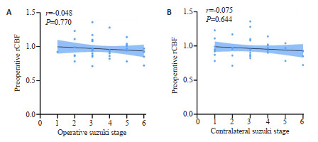
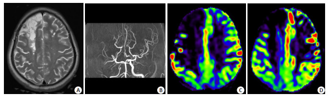
Methods: We prospectively collected the clinical and radiographic data of 40 adult patients with moyamoya disease diagnosed by digital subtraction angiography (DSA) or magnetic resonance angiography (MRA) undergoing unilateral superficial temporal artery-middle cerebral artery (STA-MCA) anastomosis. All the patients underwent 3D-pCASL examination before and after the surgery, and were followed up for 3 to 11 months after the operation. The region of interest (ROI) was located in the middle cerebral artery cortical territory covering the surgical side and ipsilateral cerebellar hemisphere. Cerebral blood flow (CBF) and relative CBF (rCBF) values were compared before and after the surgery, and the changes in cerebral hemodynamics were evaluated. The correlations were analyzed between preoperative rCBF and Suzuki stage and between the changes in postoperative neurological symptoms and rCBF.
Results: The mean CBF of the patients increased significantly from 53.96±10.04 mL·100 g-1·min-1 to 58.90±13.57 mL·100 g-1·min-1 after the operation (t=-3.068, P=0.004); the mean rCBF also increased significantly after the operation (0.96 ± 0.14 vs 1.15 ± 0.18; t=-7.155, P < 0.001). The changes in postoperative neurological symptoms were significantly correlated with the changes in rCBF (P=0.017) and the type of disease onset (P < 0.001).
Conclusion: 3D-pCASL is an valuable means for noninvasive monitoring of hemodynamic changes after revascularization in adult patients with moyamoya disease without the use of contrast agent.
目的: 利用三维伪连续动脉自旋标记(3D-pCASL)技术评估成年烟雾病患者血管重建术后脑血流动力学的变化及其与神经症状预后的关系。
方法: 收集我院经DSA或MRA确诊并接受单侧颞浅动脉(STA)-大脑中动脉(MCA)搭桥术的40例成年烟雾病患者,所有患者均于术前及术后行3D-pCASL检查,术后随访时间为3~11月。在覆盖手术侧大脑中动脉皮层区域及同侧小脑半球勾画感兴趣区(ROI),比较术前术后脑血流量(CBF)及相对脑血流量(rCBF)值,评估脑血流动力学改变情况。分析术前rCBF与铃木(Suzuki)分期的关系以及术后神经症状变化与rCBF变化之间的关系。
结果: 手术前后CBF分别为(53.96 ± 10.04)mL/100 g·min、(58.90±13.57)mL/100 g·min,差异有统计学意义(t=-3.068,P=0.004);rCBF分别0.96±0.14、1.15±0.18,差异有统计学意义(t=-7.155,P < 0.001)。术后神经症状变化与rCBF变化(P=0.017)及发病类型(P < 0.001)有关。
结论: 3D-pCASL可以用于监测成年烟雾病患者血管重建术后血流动力学变化情况,且由于其无创、无需使用对比剂等特点,值得在临床推广。
Keywords: Moyamoya disease; cerebral hemodynamics; revascularization; three-dimensional pseudo-continuous arterial spin labeling.
Figures

Similar articles
-
Arterial spin-labeling cerebral perfusion changes after revascularization surgery in pediatric moyamoya disease and syndrome.J Neurosurg Pediatr. 2019 Apr 1;23(4):486-492. doi: 10.3171/2018.11.PEDS18498. Epub 2019 Feb 8. J Neurosurg Pediatr. 2019. PMID: 30738390
-
Prediction of Cerebral Hyperperfusion after Superficial Temporal Artery-Middle Cerebral Artery Anastomosis by Three-Dimensional-Time-of-Flight Magnetic Resonance Angiography in Adult Patients with Moyamoya Disease.Cerebrovasc Dis. 2020;49(4):396-403. doi: 10.1159/000509740. Epub 2020 Aug 21. Cerebrovasc Dis. 2020. PMID: 32829323
-
Perfusion-weighted magnetic resonance imaging used in assessing hemodynamics following superficial temporal artery-middle cerebral artery bypass in patients with Moyamoya disease.Cerebrovasc Dis. 2013;35(5):455-60. doi: 10.1159/000350197. Epub 2013 May 31. Cerebrovasc Dis. 2013. PMID: 23735877 Clinical Trial.
-
Arterial Spin Labeling MRI for Quantitative Assessment of Cerebral Perfusion Before and After Cerebral Revascularization in Children with Moyamoya Disease.Korean J Radiol. 2019 Jun;20(6):985-996. doi: 10.3348/kjr.2018.0651. Korean J Radiol. 2019. PMID: 31132824 Free PMC article.
-
Quantitative hemodynamic studies in moyamoya disease: a review.Neurosurg Focus. 2009 Apr;26(4):E5. doi: 10.3171/2009.1.FOCUS08300. Neurosurg Focus. 2009. PMID: 19335131 Free PMC article. Review.
References
-
- Fujimura M, Tominaga T. Flow-augmentation bypass for moyamoya disease. J Neurosurg Sci. 2021;65(3):277–86. - PubMed
-
- Kuroda S, Nakayama N, Yamamoto S, et al. Late (5-20 years) outcomes after STA-MCA anastomosis and encephalo-duro-myoarterio-pericranial synangiosis in patients with moyamoya disease. J Neurosurg. 2020;134(3):909–16. - PubMed
-
- Fujimura M, Kaneta T, Mugikura S, et al. Temporary neurologic deterioration due to cerebral hyperperfusion after superficial temporal artery-middle cerebral artery anastomosis in patients with adult-onset moyamoya disease. Surg Neurol. 2007;67(3):273–82. doi: 10.1016/j.surneu.2006.07.017. - DOI - PubMed
-
- Fujimura M, Shimizu H, Inoue T, et al. Significance of focal cerebral hyperperfusion as a cause of transient neurologic deterioration after extracranial-intracranial bypass for moyamoya disease: comparative study with non-moyamoya patients using N-isopropyl-p-[(123)I] iodoamphetamine single-photon emission computed tomography. Neurosurgery. 2011;68(4):957–64;discussion964-5. doi: 10.1227/NEU.0b013e318208f1da. - DOI - PubMed
Publication types
MeSH terms
Substances
Supplementary concepts
LinkOut - more resources
Full Text Sources
Miscellaneous