. 2023 Apr 23;9(1):63.
doi: 10.1186/s40792-023-01644-5.

Prepancreatic postduodenal portal vein: a case report and literature review

Affiliations

Prepancreatic postduodenal portal vein: a case report and literature review

Masanori Akashi et al. Surg Case Rep. .

Abstract

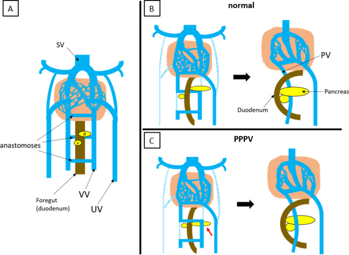
Background: Among congenital anomalies of the portal venous system, prepancreatic postduodenal portal vein (PPPV) is very rare and has only been reported to date. Herein, we report a case of PPPV identified in preoperative examinations for hepatocellular carcinoma and a literature review.

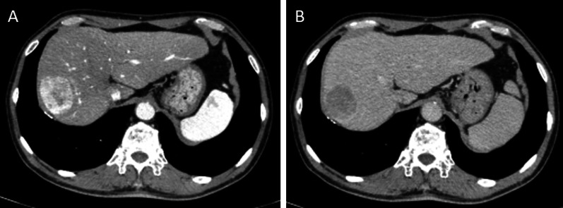
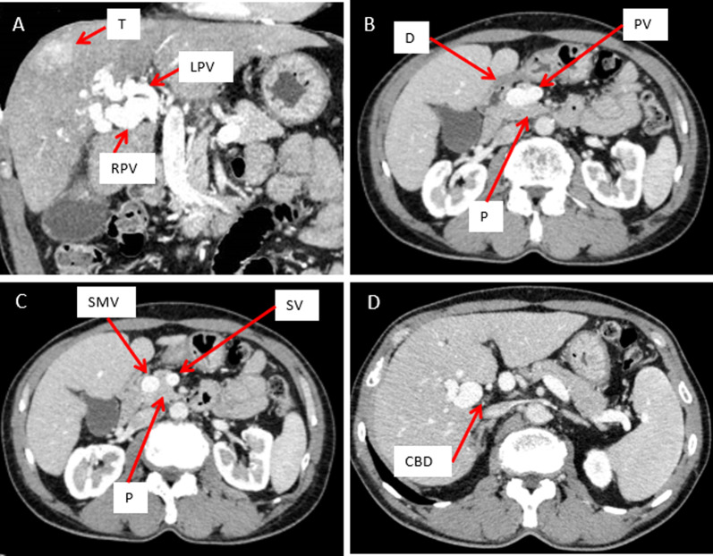
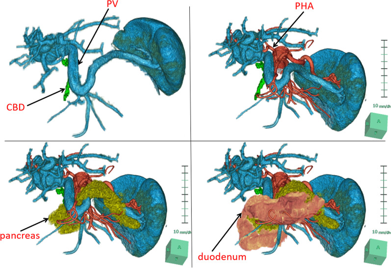
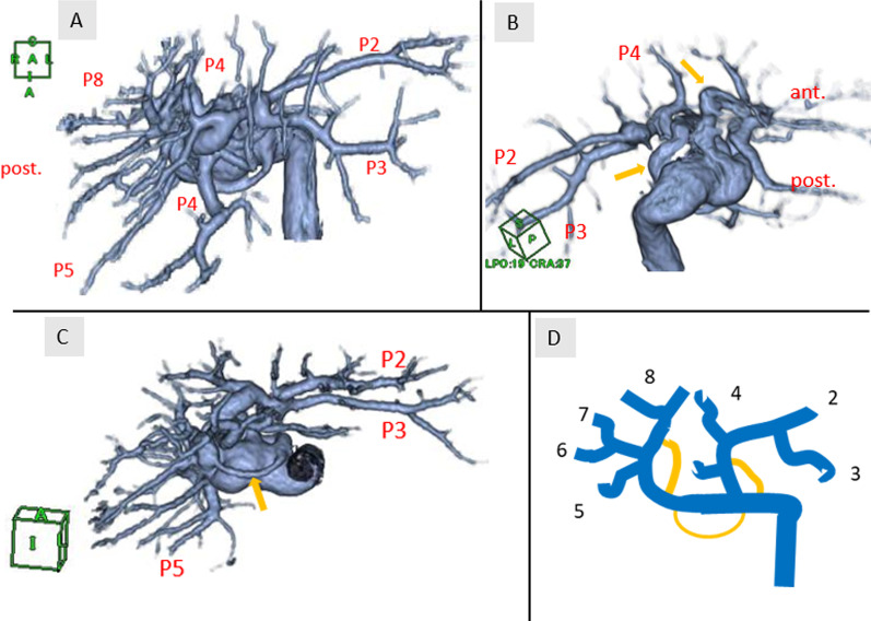
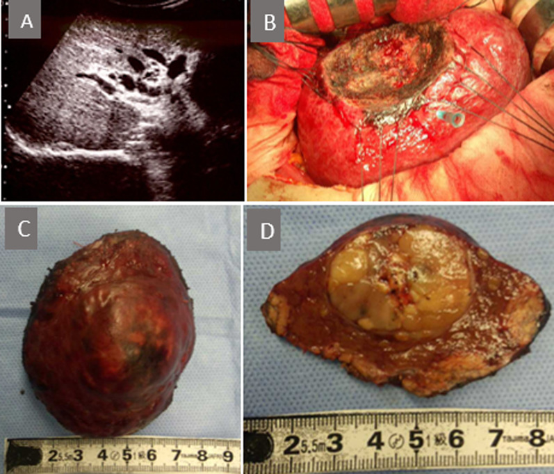
Case presentation: A 63-year-old man was admitted to our hospital for treatment of a liver tumor. After examination, he was diagnosed with hepatocellular carcinoma with a diameter of 40 mm in segment 8. Contrast-enhanced computed tomography scan showed a portal vein passing between the duodenum and pancreas, hence called PPPV. At the hepatic hilus, the portal vein branched off in a complicated course with some porto-portal communications. We determined that anatomical resection with manipulation of the hepatic hilum in this case resulted in major vascular injury. Therefore, we performed partial liver resection, and the patient was discharged uneventfully on postoperative day 14.

Conclusions: Although PPPV is an extremely rare congenital vascular variant, it is important to carefully identify vascular patterns preoperatively and to recognize the possibility of such an anomaly to avoid misidentification and inadvertent injuries during surgery.

Keywords: Anomaly of the portal venous system; Hepatectomy; Hepatocellular carcinoma; Prepancreatic postduodenal portal vein.


Conflict of interest statement

The authors declare that they have no competing interests to report.

Figures

Fig. 1
Fig. 1
Contrast-enhanced computed tomography findings (focused on tumor). A Early phase. B Delayed phase. A 40-mm tumor in segment 8 of the liver, which was enhanced in the early phase and washed out in the delayed phase, suggesting hepatocellular carcinoma
Fig. 2
Fig. 2
Contrast-enhanced computed tomography findings (focused on portal vein). A Coronal view. Portal vein dilated in the hepatic hilus and bent and twisted while branching off. B, C Portal vein was lying ventral to the pancreas and dorsal to the duodenum. D Portal vein was lying ventral to the common bile duct. T tumor, RPV right portal vein, LPV left portal vein, D duodenum, PV portal vein, P pancreas, SMV superior mesenteric vein, CBD common bile duct
Fig. 3
Fig. 3
Volume rendering 3D reconstruction of computed tomography. The image shows the portal vein running a prepancreatic postduodenal course, lying in front of the common bile duct, and forming an inverted L-shape, convexly caudad. CBD common bile duct, PV portal vein, PHA proper hepatic artery
Fig. 4
Fig. 4
AC Volume rendering 3D reconstruction of computed tomography (focused on hepatic hilus). The image shows the complex branching pattern of the portal vein and some porto-portal communications (yellow arrow). D Scheme illustrates portal vein branching and the yellow line indicates porto-portal communications
Fig. 5
Fig. 5
A Intraoperative ultrasonography showed the complex branching of the portal vein in hilus of the liver. B Liver partial resection was performed. C, D Resected specimen
Fig. 6
Fig. 6
Development of the portal vein. A Paired vitelline veins are connected by three anastomoses. The duodenum lies ventral to the intermediate anastomosis. The dorsal pancreatic bud arises ventrally to the left vitelline vein, and the ventral pancreatic bud arises contralateral and slightly caudal to the dorsal pancreatic bud. B Normal development of portal vein and pancreas. The lower part of the right vitelline vein and the upper part of the left vitelline vein disappear, and the intermediate anastomosis on the dorsal side of the duodenum forms the portal vein main trunk. As the duodenum rotates, the ventral pancreatic bud fuses behind the dorsal pancreatic bud. C Development of the prepancreatic postduodenal portal vein. The dorsal pancreatic bud arises on the dorsal side of the left vitelline vein, or arises caudal to the intermediate anastomosis, which results in the portal vein posterior to the duodenum but anterior to the pancreas. SV sinus venosus, d dorsal pancreatic bud, v ventral pancreatic bud, VV vitelline vein, UV umbilical vein, PV portal vein, PPPV prepancreatic postduodenal portal vein

References

    1. Zula DJ, Houlton AY, Nataraja RM, Pacilli M. Preduodenal portal vein associated with intestinal malrotation and jejunal atresia. Cureus. 2021;13:e16467. doi: 10.7759/cureus.16467. - DOI - PMC - PubMed
    1. Thirumoorthi AS, Cowles RA. Preduodenal portal vein. Surgery. 2016;159:672–673. doi: 10.1016/j.surg.2014.08.042. - DOI - PubMed
    1. Marks C. Developmental basis of the portal venous system. Am J Surg. 1969;117:671–681. doi: 10.1016/0002-9610(69)90404-8. - DOI - PubMed
    1. Collardeau-Frachon S, Scoazec JY. Vascular development and differentiation during human liver organogenesis. Anat Rec (Hoboken) 2008;291:614–627. doi: 10.1002/ar.20679. - DOI - PubMed
    1. Henry BM, Skinningsrud B, Saganiak K, Pękala PA, Walocha JA, Tomaszewski KA. Development of the human pancreas and its vasculature—an integrated review covering anatomical, embryological, histological, and molecular aspects. Ann Anat. 2019;221:115–124. doi: 10.1016/j.aanat.2018.09.008. - DOI - PubMed

LinkOut - more resources