. 2023 Jun:62:102705.
doi: 10.1016/j.redox.2023.102705. Epub 2023 Apr 17.

UTP11 deficiency suppresses cancer development via nucleolar stress and ferroptosis

Affiliations

UTP11 deficiency suppresses cancer development via nucleolar stress and ferroptosis

Yu Gan et al. Redox Biol. 2023 Jun.

Abstract

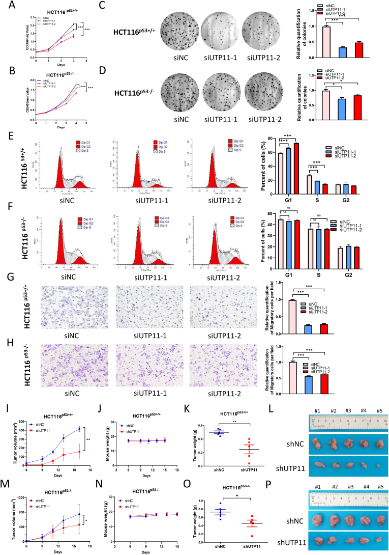
The eukaryotic ribosome is essential for cancer cell survival. Perturbation of ribosome biogenesis induces nucleolar stress or ribosomal stress, which restrains cancer growth, as rapidly proliferating cancer cells need more active ribosome biogenesis. In this study, we found that UTP11 plays an important role in the biosynthesis of 18S ribosomal RNAs (rRNA) by binding to the pre-rRNA processing factor, MPP10. UTP11 is overexpressed in human cancers and associated with poor prognoses. Interestingly, depletion of UTP11 inhibits cancer cell growth in vitro and in vivo through p53-depedednt and -independent mechanisms, whereas UTP11 overexpression promotes cancer cell growth and progression. On the one hand, the ablation of UTP11 impedes 18S rRNA biosynthesis to trigger nucleolar stress, thereby preventing MDM2-mediated p53 ubiquitination and degradation through ribosomal proteins, RPL5 and RPL11. On the other hand, UTP11 deficiency represses the expression of SLC7A11 by promoting the decay of NRF2 mRNA, resulting in reduced levels of glutathione (GSH) and enhanced ferroptosis. Altogether, our study uncovers a critical role for UTP11 in maintaining cancer cell survival and growth, as depleting UTP11 leads to p53-dependent cancer cell growth arrest and p53-independent ferroptosis.

Keywords: Ferroptosis; Nucleolar stress; Ribosome biogenesis; SLC7A11; UTP11; p53.


Conflict of interest statement

Declaration of competing interest The authors declare no competing interests.

Figures

Fig. 1
Fig. 1
Ablation of UTP11 induces p53 activation. (A) Schematic diagram depicts the screening for UTP11. (B) The heatmap of RNA-sequencing analysis of CAL-51 breast cancer cells reveals that p53 target genes are induced by UTP11 knockdown. (C) Signaling pathways that are enriched in UTP11-depleted cells are displayed by Kyoto Encyclopedia of Genes and Genomes (KEGG) analysis. (DI) Knockdown of UTP11 increases the expression of p53 and its target genes. CAL-51 (D, E), MCF-7 (F, G) and HCT116 p53+/+ cells (H, I) were transfected with control or UTP11 siRNAs, followed by IB and RT-qPCR analyses. (J, K) Knockdown of UTP11 has no effect on the expression of p53 and its target genes in HCT116p53−/ cells. ***p < 0.001.
Fig. 2
Fig. 2
UTP11 is required for 18S rRNA synthesis by binding to MPP10. (AD) Knockdown of UTP11 reduces the production of 18S rRNA. CAL-51 and HCT116 p53+/+ cells were transfected with control or UTP11 siRNA, followed by agarose gel electrophoresis (A, C) and quantification by densitometry (B, D). (EF) Knockdown of UTP11 selectively reduces levels of 18S rRNA. Fragments of 18S or 28S rRNA were detected by RT-qPCR in CAL-51 (E) and HCT116 p53+/+ cells (F) transfected with control or UTP11 siRNA. (G) Knockdown of UTP11 reduces the formation of 18S rRNA processing intermediates 21S and 18SE RNA. Cells were transfected with control or UTP11 siRNA, followed by Northern blotting analysis. A schematic illustration of 18S rRNA processing with the probe (orange) used is shown in the left panel. (H) UTP11 depletion disrupts the nucleolar localization of NPM1. Cells were transfected with control or UTP11 siRNA, followed by IF staining. (I) UTP11 and MPP10 are co-localized in the nucleolus. Cells were transfected with Flag-UTP11 and Myc-MPP10, followed by IF staining using anti-Flag and anti-Myc antibodies. (J, K) UTP11 interacts with MPP10. Cells were transfected with the indicated plasmids, followed by co-IP-IB analysis using antibodies as indicate. (L) Endogenous interaction of UTP11 and MPP10. HCT116 p53+/+ cells were treated with MG132 for 6 h and subjected to co-IP-IB analysis. ***p < 0.001. (For interpretation of the references to color in this figure legend, the reader is referred to the Web version of this article.)
Fig. 3
Fig. 3
Ablation of UTP11 stabilizes p53 through RPL5/RPL11 inhibition of MDM2. (AD) Knockdown of RPL5 or RPL11 compromises the induction of p53 by UTP11 depletion. CAL-51 (A, B) and HCT116 p53+/+ cells (C, D) were transfected with control, UTP11 siRNA, RPL5 siRNA, and RPL11 siRNA as indicated. Cell lysates were subjected to IB analysis with indicated antibodies. (E, F) RPL5-MDM2 and RPL11-MDM2 interactions are increased by depletion of UTP11. CAL-51 cells were transfected with control or UTP11 siRNA, followed by co-IP-IB assays using antibodies as indicated. The proteasome inhibitor MG132 was supplemented into medium for 5 h before cell harvest. (G) Knockdown of UTP11 diminishes MDM2-induced p53 ubiquitination. HCT116 p53−/ cells stably expressing control or UTP11 shRNA were transfected with plasmids encoding p53, His-Ub, and HA-MDM2 as indicated and treated with MG132 for 5 h, followed by in vivo ubiquitination assay and IB analysis. (H) UPT11 knockdown extends the half-life of p53 protein. CAL-51 cells were transfected with control or UTP11 siRNA. CHX (100 mg/ml) was supplemented into medium for the indicated time before cells were harvested for IB analysis. ***p < 0.001.
Fig. 4
Fig. 4
UTP11 deficiency inhibits breast cancer cell growth and migration. (A, B) Knockdown of UTP11 suppresses proliferation of wild-type p53-harboring cancer cells. CAL-51 (A) and MCF-7 cells (B) were transfected with control or UTP11 siRNAs for 6–12 h and seeded in 96-well plates for cell viability assay. (C, D) Knockdown of UTP11 inhibits clonogenic ability of wild-type p53-harboring cancer cells. CAL-51 (C) and MCF-7 cells (D) were seeded in 6-well plates for about 14 days. Colonies were fixed with methanol, and visualized by crystal violet staining. (E, F) Knockdown of UTP11 induces G1 cell cycle arrest in wild-type p53-harboring cancer cells. CAL-51 (E) and MCF-7 cells (F) were transfected with control or UTP11 siRNAs, followed by flow cytometry analysis. (G, H) Knockdown of UTP11 impede migration of wild-type p53-harboring cancer cells. CAL-51 (G) and MCF-7 cells (H) were transfected with control or UTP11 siRNAs, followed by transwell cell migration assay. (IL) UTP11 deficiency suppresses cancer growth in vivo. Depletion of UTP11 suppresses CAL-51 cell-derived xenograft tumor growth rate (I), weight (K), and size (L), while has no effect on mouse body weight (J). The data are represented as mean ± SD, n = 10. ***p < 0.001. (For interpretation of the references to color in this figure legend, the reader is referred to the Web version of this article.)
Fig. 5
Fig. 5
UTP11 deficiency-mediated tumor-inhibitory effects are partially dependent on p53. (A, B) Knockdown of UTP11 restrains proliferation of HCT116p53+/+ cells (A) more dramatically than HCT116 p53−/ cells (B). Cells were transfected with control or UTP11 siRNAs for 6–12 h and seeded in 96-well plates, followed by cell viability assay. (C, D) UTP11 depletion reduces the colony-forming ability of HCT116 p53+/+ cells (C) more dramatically than HCT116 p53−/ cells (D). Cells were seeded in 6-well plates for about 14 days. Colonies were fixed with methanol, and visualized by crystal violet staining. (E, F) UTP11 deficiency induces G1 arrest in HCT116 p53+/+ (E) but not HCT116 p53−/−cells (F). Cells were transfected with control or UTP11 siRNAs, followed by flow cytometry analysis. (G, H) Knockdown of UTP11 impedes migration of HCT116 p53+/+ cells (G) more dramatically than HCT116 p53−/ cells (H). (I–P) UTP11 depletion suppresses tumor growth partially dependently on p53 in vivo. UTP11 depletion dramatically suppresses HCT116 p53+/+ cell-derived tumor growth rate (I), weight (K), and size (L), while also moderately reduces HCT116 p53−/ cell-derived tumor growth rate (M), weight (O), and size (P). Mouse body weight was not affected (J, N). The data are represented as mean ± SD, n = 5. *p < 0.05, **p < 0.01, ***p < 0.001. (For interpretation of the references to color in this figure legend, the reader is referred to the Web version of this article.)
Fig. 6
Fig. 6
UTP11 deficiency triggers ferroptosis by regulating the NRF2-SLC7A11 axis. (A-D) Knockdown of UTP11 represses SLC7A11 mRNA and protein expression independently of p53. HCT116 p53+/+ (A, B) and HCT116 p53−/ cells (C, D) transfected with the indicated siRNAs were subjected to RT-qPCR and IB analyses. (E, F) UTP11 depletion reduces GSH levels. HCT116 p53+/+ (E) and HCT116 p53−/- cells (F) were transfected with the indicated siRNAs, followed by GSH assay. (G, H) UTP11 depletion elevates MDA levels. HCT116 p53+/+ (G) and HCT116 p53−/- cells (H) were transfected with the indicated siRNAs, followed by MDA assay. (I, J) UTP11 depletion-caused cell growth inhibition is partially restored by Ferrostatin-1. HCT116 p53+/+ (I) and HCT116p53−/ cells (J) transfected with the indicated siRNAs were seeded in 96-well plates and treated with DMSO or Ferrostatin-1 (2 μM) for 48 h, followed by cell viability assay. (K) RNA-sequencing results reveal that knockdown of UTP11 reduces the expression of NRF2 and its target genes. (LO) UTP11 depletion markedly represses NRF2 and its target gene expression. HCT116 p53+/+ (L, M) and HCT116 p53−/- cells (N, O) transfected with the indicated siRNAs were subjected to RT-qPCR and IB analyses. (PR) UTP11 interacts with NRF2 mRNA. HCT116 p53−/- cells were transfected with control or Flag-UTP11, and input samples were confirmed by IB (P). RNA immunoprecipitation (RIP) assays were performed to detect the interactions between UTP11 and NRF2 mRNA, followed by RT-qPCR (Q) and agarose gel electrophoresis (R). Two pairs of primers were designed to amplify fragments from 448 to 617 bp and 1048–1275 bp of NRF2 mRNA. (S) UTP11 depletion decreases NRF2 mRNA stability. HCT116 p53−/- cells transfected with control or UTP11 siRNA were treated with actinomycin D (5 μg/ml) for the indicated time, followed by RT-qPCR analysis. (T, U) UTP11 deficiency prevents NRF2 recruitment on SLC7A11 promoter. HCT116 p53−/- cells were transfected with siRNAs or plasmids as indicated, followed by ChIP- qPCR (T) and IB (U) analyses. *p < 0.05, **p < 0.01, ***p < 0.001.
Fig. 7
Fig. 7
UTP11 overexpression is correlated with unfavorable prognoses in breast and colorectal cancers (A) UTP11 protein expression is upregulated in breast cancer samples compared to adjacent normal tissues. Six pairs of tissues were analyzed by IB. (B) UTP11 mRNA expression is upregulated in breast cancer samples compared to adjacent normal tissues. Ten pairs of tissues were analyzed by RT-qPCR. (C, D) Higher expression of UTP11 is significantly associated with shorter overall survival in a cohort of 91 breast cancer patients. (E, F) UTP11 protein expression is higher in colorectal cancer samples than adjacent normal tissues. Representative images (E) and the graph of ten pairs of samples (F) are shown. (G, H) Higher levels of UTP11 are associated with worse prognoses in 150 colorectal cancer patients. (I) A schematic diagram for UTP11 function in cancer. In UTP11-proficient cells, p53 is degraded through MDM2-mediated ubiquitination and lipid ROS is restricted via NRF2 antioxidant signaling pathway (e.g., SLC7A11), which promotes cancer cell survival and growth (left panel). In UTP11-deficient cells, the biosynthesis of 18S rRNA is impaired, resulting in nucleolar stress-induced p53 stabilization and activation. Additionally, UTP11 depletion promotes the destabilization of NRF2 mRNA, leading to the downregulation of SLC7A11 and increased ferroptosis (right panel). **p < 0.01.

References

    1. Pelletier J., Thomas G., Volarevic S. Ribosome biogenesis in cancer: new players and therapeutic avenues. Nat. Rev. Cancer. 2018;18(1):51–63. - PubMed
    1. Zhou X., Lu H. In: Encyclopedia of Cell Biology. Bradshaw R., Hart G., Stahl P., editors. Academic Press; 2023. Extra-ribosome functions of ribosomal proteins; pp. 57–70.
    1. van Riggelen J., Yetil A., Felsher D.W. MYC as a regulator of ribosome biogenesis and protein synthesis. Nat. Rev. Cancer. 2010;10(4):301–309. - PubMed
    1. Zhai W., Comai L. Repression of RNA polymerase I transcription by the tumor suppressor p53. Mol. Cell Biol. 2000;20(16):5930–5938. - PMC - PubMed
    1. Chesnokov I., et al. p53 inhibits RNA polymerase III-directed transcription in a promoter-dependent manner. Mol. Cell Biol. 1996;16(12):7084–7088. - PMC - PubMed

Publication types

MeSH terms

Substances