. 2023 Jun;22(6):100557.
doi: 10.1016/j.mcpro.2023.100557. Epub 2023 Apr 22.

Ultrasensitive Protein Detection Technologies for Extracellular Vesicle Measurements

Affiliations

Ultrasensitive Protein Detection Technologies for Extracellular Vesicle Measurements

Adnan Shami-Shah et al. Mol Cell Proteomics. 2023 Jun.

Abstract

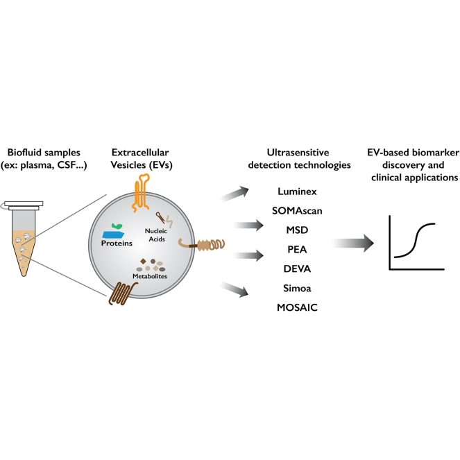
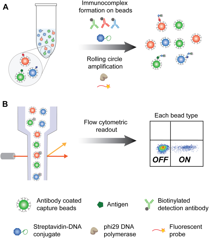
Extracellular vesicles (EVs) are nanoscopic, heterogenous, lipid-rich particles that carry a multitude of cargo biomolecules including proteins, nucleic acids, and metabolites. Although historically EVs were regarded as cellular debris with no intrinsic value, growing understanding of EV biogenesis has led to the realization that EVs facilitate intercellular communication and are sources of liquid biomarkers. EVs can be isolated and analyzed from a wide variety of accessible biofluids for biomarker discovery and diagnostic applications. There is a diversity of EVs from different biological compartments (e.g., cells and tissues), and some of these EVs are present at extremely low concentrations. Consequently, a challenge in the field is to find appropriate markers that enable selective isolation of these rare EVs. Many conventional protein detection technologies have limited sensitivity to detect low abundance biomarkers in EVs, limiting their use in EV research. Advances in ultrasensitive detection technologies are needed to harness the potential of EVs for clinical application. This Perspective highlights current EV research focusing on ultrasensitive detection technologies, their limitations, and areas of potential growth in the future.

Keywords: biofluids; biomarkers; extracellular vesicles; proteins; ultrasensitive technology.


Conflict of interest statement

Conflict of interest The authors declare the following financial interests/personal relationships which may be considered as potential competing interests: A. S. and M. N. declare no known competing financial interests. D. R. W. has a financial interest in Quanterix, a company developing an ultrasensitive digital immunoassay platform; he is an inventor of the Simoa technology, a founder of the company, and a member of its board of directors. D. R. W.’s interests were reviewed and are managed by BWH. and Partners HealthCare in accordance with their conflict-of-interest policies.

Figures

None
Graphical abstract
Fig. 1
Fig. 1
Schematic of EV separation techniques that can be used to validate immunocapture targets.A, Size exclusion chromatography separates soluble proteins (depicted in blue) and EVs (depicted in green) by size. Most commonly a Sepharose resin is used. The EVs elute in the early fractions while free proteins elute in the late fractions. B, Density gradient chromatography separates soluble proteins and EVs based on density. Most commonly, an iodixanol gradient is used. After ultracentrifugation, the free proteins appear at the top of the resin while the EVs descend to the bottom of the resin.
Fig. 2
Fig. 2
Schematic of proteinase protection assay. The EV membrane protects the internal EV contents from proteinase until Triton X permeabilizes the membrane. In this protocol, the proteinase inhibitor PMSF is added after proteinase treatment but before Triton X to preserve the internal proteins, after all external proteins are digested. A, without treatment, both internal EV proteins and external free proteins are present. B, without proteinase treatment but with PMSF, both internal EV proteins and external free proteins are present. C, with proteinase treatment but without PMSF, both internal EV proteins and external free proteins are digested. D, with proteinase treatment and with PMSF, internal EV proteins are preserved but external free proteins are digested. When samples are being measured for internal EV protein detection, the protocol in panel d should be utilized, while (AC) serve as controls. PMSF, phenylmethylsulfonyl fluoride.
Fig. 3
Fig. 3
Schematic of Simoa assay workflow.A, target protein molecules are captured on antibody-coated capture beads. Immunocomplex is then formed on the beads by labeling the protein with a biotinylated detection antibody and streptavidin-ß-galactosidase. B, individual beads are loaded onto a microarray of femtoliter-sized wells such that only one bead can fit into a well. The microarray is then sealed with silicone oil and fluorescence images of the array are acquired. Simoa, Single-molecule array.
Fig. 4
Fig. 4
Schematic of MOSAIC assay workflow.A, target protein molecules are captured with an excess number of antibody-coated capture beads. Immunocomplexes, formed on the beads by labeling with biotinylated detection antibody and streptavidin-DNA conjugate, undergo rolling circle amplification (RCA) to form a long DNA concatemer. Fluorescently labeled DNA probes are then hybridized to the RCA product. B, flow cytometry is used to count on and off beads for each bead type. MOSAIC, Molecular On-bead Signal Amplification for Individual Counting.

Similar articles

Cited by

References

    1. Salomon C., Das S., Erdbrügger U., Kalluri R., Lim S.K., Olefsky J.M., et al. Extracellular vesicles and their emerging roles as cellular messengers in endocrinology: an endocrine society scientific statement. Endocr. Rev. 2022;43:441–468. - PMC - PubMed
    1. Doyle L., Wang M.Z. Overview of extracellular vesicles, their origin, composition, purpose, and methods for exosome isolation and analysis. Cells. 2019;8:41–68. - PMC - PubMed
    1. Wolf P. The nature and significance of platelet products in human plasma. Br. J. Haematol. 1967;13:269–288. - PubMed
    1. Shao H., Im H., Castro C.M., Breakefield X., Weissleder R., Lee H. New technologies for analysis of extracellular vesicles. Chem. Rev. 2018;118:1917–1950. - PMC - PubMed
    1. Chang W., Cerione R.A., Antonyak M.A. Extracellular vesicles and their roles in cancer progression. Methods Mol. Biol. 2021;2174:143–170. - PMC - PubMed

MeSH terms

LinkOut - more resources