. 2023 Apr 24;13(1):57.
doi: 10.1038/s41408-023-00830-w.

Combined inhibition of BCL-2 and MCL-1 overcomes BAX deficiency-mediated resistance of TP53-mutant acute myeloid leukemia to individual BH3 mimetics

Affiliations

Combined inhibition of BCL-2 and MCL-1 overcomes BAX deficiency-mediated resistance of TP53-mutant acute myeloid leukemia to individual BH3 mimetics

Bing Z Carter et al. Blood Cancer J. .

Erratum in

Abstract

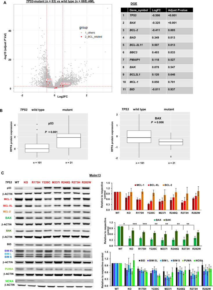
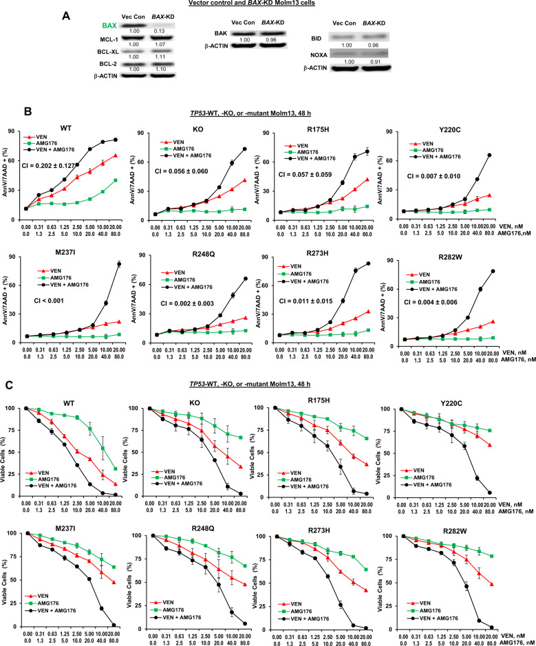
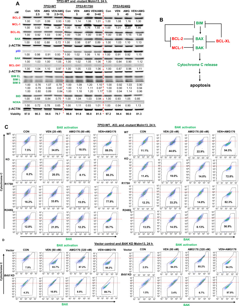
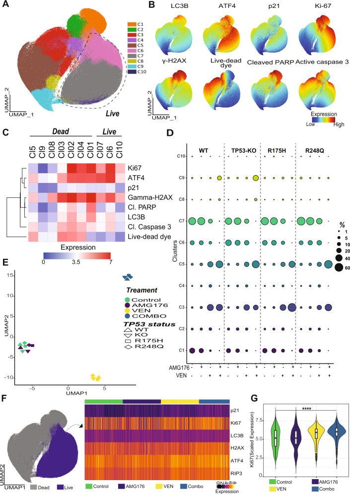
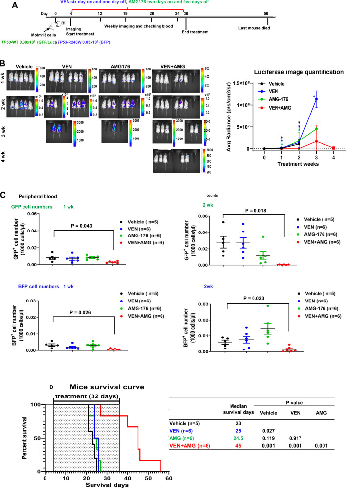
TP53-mutant acute myeloid leukemia (AML) respond poorly to currently available treatments, including venetoclax-based drug combinations and pose a major therapeutic challenge. Analyses of RNA sequencing and reverse phase protein array datasets revealed significantly lower BAX RNA and protein levels in TP53-mutant compared to TP53-wild-type (WT) AML, a finding confirmed in isogenic CRISPR-generated TP53-knockout and -mutant AML. The response to either BCL-2 (venetoclax) or MCL-1 (AMG176) inhibition was BAX-dependent and much reduced in TP53-mutant compared to TP53-WT cells, while the combination of two BH3 mimetics effectively activated BAX, circumventing survival mechanisms in cells treated with either BH3 mimetic, and synergistically induced cell death in TP53-mutant AML and stem/progenitor cells. The BH3 mimetic-driven stress response and cell death patterns after dual inhibition were largely independent of TP53 status and affected by apoptosis induction. Co-targeting, but not individual targeting of BCL-2 and MCL-1 in mice xenografted with TP53-WT and TP53-R248W Molm13 cells suppressed both TP53-WT and TP53-mutant cell growth and significantly prolonged survival. Our results demonstrate that co-targeting BCL-2 and MCL-1 overcomes BAX deficiency-mediated resistance to individual BH3 mimetics in TP53-mutant cells, thus shifting cell fate from survival to death in TP53-deficient and -mutant AML. This concept warrants clinical evaluation.


Conflict of interest statement

PEH and PKM are employees of Amgen.

Figures

Fig. 1
Fig. 1. BAX expression is significantly decreased in TP53-KO and -mutant AML cells.
A Volcano plot showing differential gene expression (DGE) and the adjusted P values of BCL-2 family members between TP53-mutant and TP53-WT AML cells, as assessed by RNA-seq analysis. DGE and adjusted P values were calculated using the limma package. TP53 and differentially expressed BCL-2–related genes are highlighted in red. B RPPA analysis of the log2-normalized expression levels of p53 and BAX in newly diagnosed de novo TP53-mutant and -WT AML. Box is 75 to 25% and bar in the middle is the median of expression. The whiskers are 95% CI, with the extremes (black dots) outside that. C Expression of BCL-2 family proteins in TP53-KO and -mutant Molm13 cells and isogenic WT control Molm13 cells. Left, a representative Western blot; right, the relative protein expression levels quantified from three Western blots. Error bars represent the mean ± SEM of three independent experiments. Statistical analysis was performed using a Student t-test, and P < 0.05 was considered statistically significant. *P < 0.05; **P < 0.01; ***P < 0.001.
Fig. 2
Fig. 2. Combined inhibition of BCL-2 and MCL-1 synergistically induces cell death and decreases cell viability in TP53-KO and -mutant cells.
A Western blotting of BCL-2 family proteins in vector control (Vec Con) and BAX-KD Molm13 cells. B, C TP53-WT, -KO, and -mutant Molm13 cells were treated with VEN, AMG176, or both for 48 h. Cell death (B) and cell viability (C) in the WT control cells and the TP53-KO and TP53-mutant cells were determined by flow cytometry. Experiments were performed in triplicate. Error bars represent the mean ± SEM of three independent experiments. h hour, AnnV annexin V, 7AAD 7-aminoactinomycin D.
Fig. 3
Fig. 3. The role of BCL-2 family members in BH3 mimetic–induced apoptosis.
A TP53-WT and -mutant (R175H and R248Q) Molm13 cells were treated with VEN, AMG176 (AMG), or both at the indicated concentrations for 24 h. Protein levels were determined by Western blot analysis. B Schematic of apoptosis induction requiring BAX and BAK activation, which is activated by BIM and blocked by BCL-2 and MCL-1. C, D TP53-WT, -KO, or -mutant Molm13 cells (C) and vector control or BAX-KD Molm13 cells (D) were treated with VEN, AMG176, or both at the indicated concentrations for 24 h. BAX and BAK activation and cytochrome C release were measured by flow cytometry. h hour.
Fig. 4
Fig. 4. The combination of VEN and AMG176 induces similar cell death and stress patterns in TP53-WT, -KO, and -mutant AML cells.
A TP53-WT, -KO, and -mutant (R175H and R248Q) Molm13 cells were treated with DMSO control, VEN, AMG176, or VEN plus AMG176, stained with an array of antibodies, and subjected to flow cytometry. Cells were then subjected to UMAP dimension reduction and projected on two-dimensional plots. The FlowSOM algorithm was utilized for automated cell population identification; the colors indicate FlowSOM clusters. B The UMAP plots for the indicated markers; the colors indicate marker expression intensity. C Heatmap of the arcsine-transformed expression intensities of the markers used for dimension reduction and clustering across the FlowSOM clusters shown in panel B. Live and dead cell clusters were identified based on the marker expression patterns shown in panel B. D Bubble plot of the frequencies of the FlowSOM clusters shown in panel A across TP53-WT, -KO, and -mutant (R175H and R248Q) Molm13 cells treated with control, VEN, AMG176, or VEN plus AMG176. E UMAP plot of the differential responses to treatment with control, VEN, AMG176, or VEN plus AMG176. The FlowSOM cluster frequencies shown in panel C were used for dimension reduction. F UMAP map of live cells (purple) and dead cells (gray) and heatmap of the expressions of the selected markers. G Violin plot of scaled Ki-67 signal intensities of TP53-WT, KO, and mutant leukemia cells and cells exposed to VEN, AMG176, or VEN plus AMG176 for 48 h. Live cells were selected for plotting and 200 cells (50 cells each of TP53-WT, KO, R175H, and R248Q) from each condition (control, AMG176, VEN, and combo) were shown. Cl cleaved, combo combination.
Fig. 5
Fig. 5. The combined inhibition of BCL-2 and MCL-1 is significantly more effective in AML cells and AML stem/progenitor cells from patients with TP53 mutations.
A, B AML patient samples (n = 8) with TP53 mutations were treated with VEN, AMG176, or both for 48 h. Apoptosis (A) and viable cells (B) were determined by flow cytometry. h hour. Error bars represent mean ± SEM of data from individual patients. C AML patient samples (n = 8) with TP53 mutations were treated with various concentrations of VEN, AMG176, or both for 48 h. Apoptosis and the CI values from two representative patient samples (#26, #28) are shown. Cell death is expressed as specific apoptosis and viable cells are expressed as % compared to the control of 100%. con control.
Fig. 6
Fig. 6. The combined inhibition of BCL-2 and MCL-1 exhibits stronger in vivo anti-leukemia activity in a xenograft mouse model.
A Experimental scheme. B Bioluminescence imaging and quantification. C Circulating numbers of GFP+ and BFP+ cells after week 1 (left) and week 2 (right) of treatment as determined by flow cytometry. D Survival. n = 5 or 6/group. AMG AMG176, wk week. Error bars represent the mean ± SD of data from individual mice.

References

    1. Christiansen DH, Andersen MK, Pedersen-Bjergaard J. Mutations with loss of heterozygosity of p53 are common in therapy-related myelodysplasia and acute myeloid leukemia after exposure to alkylating agents and significantly associated with deletion or loss of 5q, a complex karyotype, and a poor prognosis. J Clin Oncol. 2001;19:1405–13. doi: 10.1200/JCO.2001.19.5.1405. - DOI - PubMed
    1. Ok CY, Patel KP, Garcia-Manero G, Routbort MJ, Fu B, Tang G, et al. Mutational profiling of therapy-related myelodysplastic syndromes and acute myeloid leukemia by next generation sequencing, a comparison with de novo diseases. Leuk Res. 2015;39:348–54. doi: 10.1016/j.leukres.2014.12.006. - DOI - PMC - PubMed
    1. Pedersen-Bjergaard J, Andersen MK, Andersen MT, Christiansen DH. Genetics of therapy-related myelodysplasia and acute myeloid leukemia. Leukemia. 2008;22:240–8. doi: 10.1038/sj.leu.2405078. - DOI - PubMed
    1. Rücker FG, Schlenk RF, Bullinger L, Kayser S, Teleanu V, Kett H, et al. TP53 alterations in acute myeloid leukemia with complex karyotype correlate with specific copy number alterations, monosomal karyotype, and dismal outcome. Blood. 2012;119:2114–21. doi: 10.1182/blood-2011-08-375758. - DOI - PubMed
    1. Hou HA, Chou WC, Kuo YY, Liu CY, Lin LI, Tseng MH, et al. TP53 mutations in de novo acute myeloid leukemia patients: longitudinal follow-ups show the mutation is stable during disease evolution. Blood Cancer J. 2015;5:e331. doi: 10.1038/bcj.2015.59. - DOI - PMC - PubMed

Publication types

MeSH terms