

A canine thromboembolic model of anterior circulation large vessel occlusion stroke
- PMID: 37089293
- PMCID: PMC10119506
- DOI: 10.1016/j.heliyon.2023.e14692
A canine thromboembolic model of anterior circulation large vessel occlusion stroke
Abstract
Purpose: To develop a large animal preclinical model of thromboembolic stroke with stable, protracted large vessel occlusion (LVO) utilizing an autologous clot.
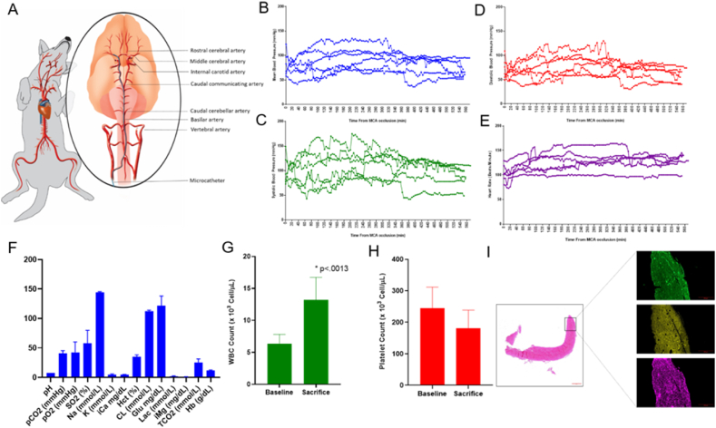
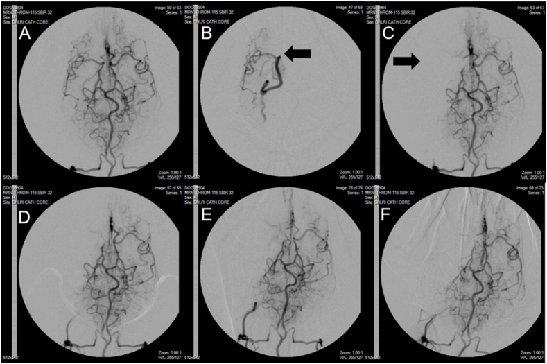
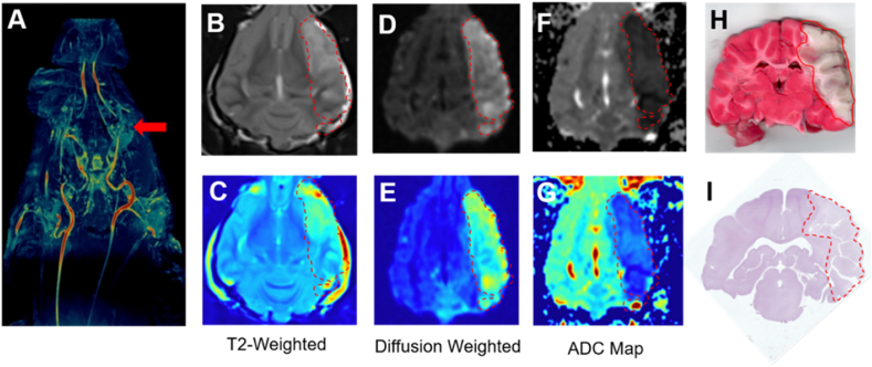
Materials and methods: A reproducible canine model of large vessel occlusion stroke was established by endovascular placement of an autologous clot into the middle cerebral artery (MCA) of six adult hounds and confirmed using digital subtraction angiography (DSA). Infarct volume and evidence of hemorrhage were determined by magnetic resonance imaging (MRI) 7 h after occlusion and Thrombolysis in Cerebral Infarction scale (TICI) was assessed before and after clot placement and at 1, 6, 7, and 9 h after middle cerebral artery occlusion (MCAO). Heart rate (HR) and blood pressure (BP) were monitored continuously and invasively through an arterial sheath throughout the procedures and complete blood count and blood gas analysis completed at time of sacrifice. Histopathological findings at time of sacrifice were used to confirm stroke volume and hemorrhage.
Results: MCAO with resulting TICI 0 flow was observed in all six animals, verified by serial DSA, and lack of collateral flow persisted for 9 h after clot placement until time of sacrifice. The mean infarct volume was 47.0 ± 6.7% of the ipsilateral hemisphere and no events of spontaneous recanalization or clot autolysis were observed.
Conclusion: We demonstrate a thromboembolic canine model of MCAO that is both feasible and results in consistent infarct volumes to generate a clinically relevant LVO. This model is important to evaluate treatment of LVO in acute ischemic stroke (AIS) outside the established 4.5 h recombinant tissue plasminogen activator (rTPA) therapeutic window utilizing a prolonged occlusive thrombus.
Keywords: Canine model; Middle cerebral artery occlusion; Stroke.
©2023PublishedbyElsevierLtd.
Conflict of interest statement
The authors declare that they have no known competing financial interests or personal relationships that could have appeared to influence the work reported in this paper.
Figures
Similar articles
-
Von Willebrand factor targeted thrombolysis in canine basilar artery occlusion.Front Neurol. 2024 Oct 9;15:1436291. doi: 10.3389/fneur.2024.1436291. eCollection 2024. Front Neurol. 2024. PMID: 39445200 Free PMC article.
-
Severe Cerebral Small Vessel Disease Burden Is Associated With Poor Outcomes After Endovascular Thrombectomy in Acute Ischemic Stroke With Large Vessel Occlusion.Cureus. 2021 Feb 4;13(2):e13122. doi: 10.7759/cureus.13122. Cureus. 2021. PMID: 33728139 Free PMC article.
-
Absence of Collaterals is Associated with Larger Infarct Volume and Worse Outcome in Patients with Large Vessel Occlusion and Mild Symptoms.J Stroke Cerebrovasc Dis. 2019 Jul;28(7):1987-1992. doi: 10.1016/j.jstrokecerebrovasdis.2019.03.032. Epub 2019 Apr 26. J Stroke Cerebrovasc Dis. 2019. PMID: 31036341
-
Revascularization of tandem occlusions in acute ischemic stroke: review of the literature and illustrative case.Neurosurg Focus. 2017 Apr;42(4):E15. doi: 10.3171/2017.1.FOCUS16521. Neurosurg Focus. 2017. PMID: 28366063 Review.
-
A novel mouse model of thromboembolic stroke.J Neurosci Methods. 2015 Dec 30;256:203-11. doi: 10.1016/j.jneumeth.2015.09.013. Epub 2015 Sep 18. J Neurosci Methods. 2015. PMID: 26386284 Free PMC article. Review.
Cited by
-
Von Willebrand factor targeted thrombolysis in canine basilar artery occlusion.Front Neurol. 2024 Oct 9;15:1436291. doi: 10.3389/fneur.2024.1436291. eCollection 2024. Front Neurol. 2024. PMID: 39445200 Free PMC article.
References
-
- GBD 2016 Risk Factors Collaborators Global, regional, and national comparative risk assessment of 84 behavioural, environmental and occupational, and metabolic risks or clusters of risks, 1990-2016: a systematic analysis for the Global Burden of Disease Study 2016. Lancet. 2017 Sep 16;390(10100):1345–1422. Erratum in: Lancet. 2017 Oct 14;390(10104):1736. Erratum in: Lancet. 2017 Oct 28;390(10106):e38. - PMC - PubMed
-
- Kaiser E.E., West F.D. Large animal ischemic stroke models: replicating human stroke pathophysiology. Neural Regener. Res. 2020 Aug;15(8):1377–1387. Gillilan LA. Extra- and intra-cranial blood supply to brains of dog and cat. Am J Anat 1976;146(3):237-53.[published Online First: 1976/07/01] - PMC - PubMed
Grants and funding
LinkOut - more resources
Full Text Sources
