

doi: 10.4103/idoj.idoj_276_21.
eCollection 2023 Mar-Apr.
Dermoscopy of Cylindroma in Fitzpatrick Type IV Skin: A Case Report and Review of Literature
Affiliations
- PMID: 37089843
- PMCID: PMC10115342
- DOI: 10.4103/idoj.idoj_276_21
Item in Clipboard
Dermoscopy of Cylindroma in Fitzpatrick Type IV Skin: A Case Report and Review of Literature
Indian Dermatol Online J.
.
No abstract available
Conflict of interest statement
There are no conflicts of interest.
Figures
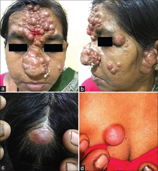
(a and b) Multiple pinkish to skin-colored discrete and coalescent nodules involving the forehead, nose, ears, and cheeks. Solitary pinkish nodules over the (c) scalp and (d) chest
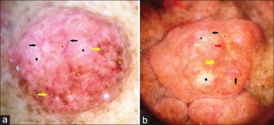
Polarized dermoscopy shows predominantly peripheral linear serpentine and branching vessels (a–b, black arrows), white structureless areas (a–b, black stars), yellow-brown structureless areas (a–b, red stars), and brown structures in the form of lines (a–b, yellow arrows), network pattern (b, red arrow) and structureless areas (a–b, yellow stars) over a pink-white background. [×10]
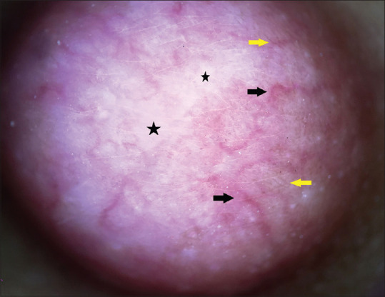
Polarized dermoscopy of the lesion on the chest reveals peripheral linear serpentine branching vessels (black arrows), white structureless areas (black stars), and thin brown lines (yellow arrows) over a pink-white background [×10]
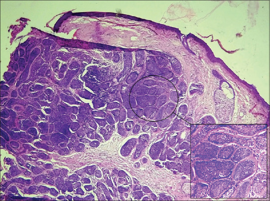
Histopathology shows multiple dermal aggregates of cellular nests composed of basaloid cells. Also seen is the hyaline eosinophilic matrix dispersed in between and around the cellular nests (inset) [H and E, ×10; inset, ×40]
Similar articles
-
Dermoscopy of cylindroma.Dermatol Res Pract. 2010;2010:285392. doi: 10.1155/2010/285392. Epub 2010 Aug 24. Dermatol Res Pract. 2010. PMID: 20862372 Free PMC article.
-
Dermoscopy of solitary cylindroma.Eur J Dermatol. 2011 Jul-Aug;21(4):645-6. doi: 10.1684/ejd.2011.1413. Eur J Dermatol. 2011. PMID: 21697054 No abstract available.
-
Dermoscopy of Localized Darier's Disease in Fitzpatrick Type IV Skin.Indian Dermatol Online J. 2020 Mar 9;11(2):298-300. doi: 10.4103/idoj.IDOJ_412_18. eCollection 2020 Mar-Apr. Indian Dermatol Online J. 2020. PMID: 32478011 Free PMC article. No abstract available.
-
Dermoscopy and ultrosound monitoring actinic keratosis with cutaneous squamous cell carcinoma: A case report and literature review.Photodiagnosis Photodyn Ther. 2022 Mar;37:102709. doi: 10.1016/j.pdpdt.2021.102709. Epub 2021 Dec 30. Photodiagnosis Photodyn Ther. 2022. PMID: 34973428 Review.
-
[Dermoscopy of nonneoplastic diseases in dark skin].Dermatologie (Heidelb). 2023 Apr;74(4):250-255. doi: 10.1007/s00105-023-05121-w. Epub 2023 Mar 1. Dermatologie (Heidelb). 2023. PMID: 36859732 Free PMC article. Review. German.
Cited by
-
Dermoscopy of Adnexal Tumors in Skin of Colour as a Diagnostic Challenge Extended to Dark Skin Tones.Cureus. 2025 Jun 21;17(6):e86501. doi: 10.7759/cureus.86501. eCollection 2025 Jun. Cureus. 2025. PMID: 40693080 Free PMC article.
References
-
- McCalmont TH, Pincus LB. Adnexal neoplasms. In: Bolognia JL, Schaffer JV, Creeoni L, editors. Dermatology. 4th ed. London: Elsevier; 2018. pp. 1930–53.
-
- Wick MR, Barnhill RL. Sweat gland tumors. In: Barnhill RL, Crowson AN, Magro CM, Pipekorn MW, editors. Dermatopathology. 3rd ed. New York: McGraw Hill companies, Inc; 2010. pp. 725–65.
-
- Senarega A, Flores L, Innocenti AC, Parra V. Dermoscopic Features of Spiradenocylindroma. Actas Dermosifiliogr. 2019;110:604–6. - PubMed
LinkOut - more resources
Full Text Sources
