

Sterol regulatory element binding transcription factor 1 promotes proliferation and migration in head and neck squamous cell carcinoma
- PMID: 37090107
- PMCID: PMC10117388
- DOI: 10.7717/peerj.15203
Sterol regulatory element binding transcription factor 1 promotes proliferation and migration in head and neck squamous cell carcinoma
Abstract
Background: Sterol-regulatory element-binding protein 1 (SREBP1) is a transcription factor involved in lipid metabolism that is encoded by sterol regulatory element binding transcription factor 1(SREBF1). SREBP1 overexpression is associated with the progression of several human tumors; however, the role of SREBP1 in head and neck squamous cell carcinoma (HNSC) remains unclear.
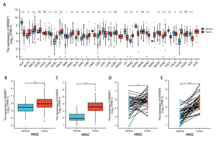
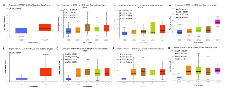
Methods: SREBF1 expression in pan-cancer was analyzed using the Cancer Genome Atlas (TCGA) and Genotype-Tissue Expression (GTEx) data, and the association between SREBF1 expression and clinical characteristics of HNSC patients was examined using the UALCAN database. Enrichment analysis of SREBF1-related genes was performed using the Cluster Profiler R package. TCGA database was used to investigate the relationship between immune cell infiltration and SREBF1 expression. CCK-8, flow cytometry, and wound healing assays were performed to investigate the effect of SREBF1 knockdown on the proliferation and migration of HNSC cells.
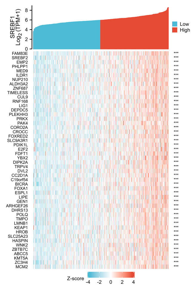
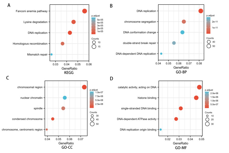
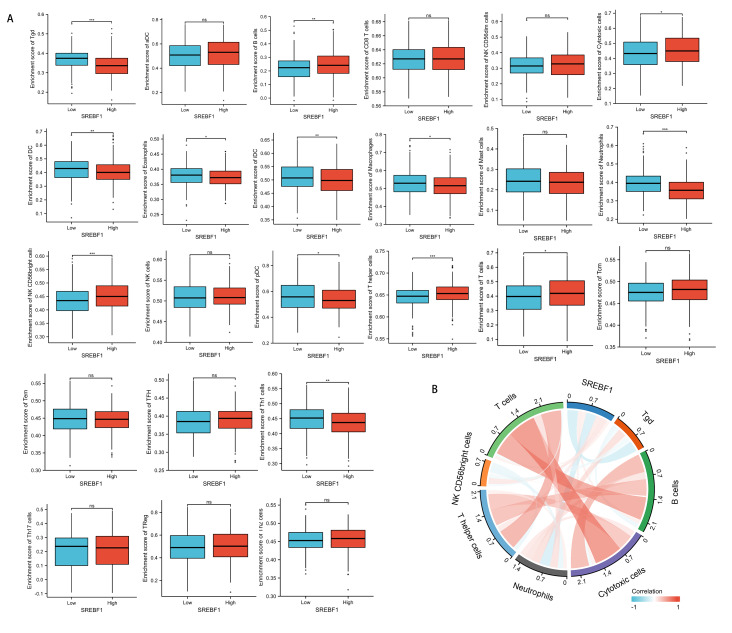
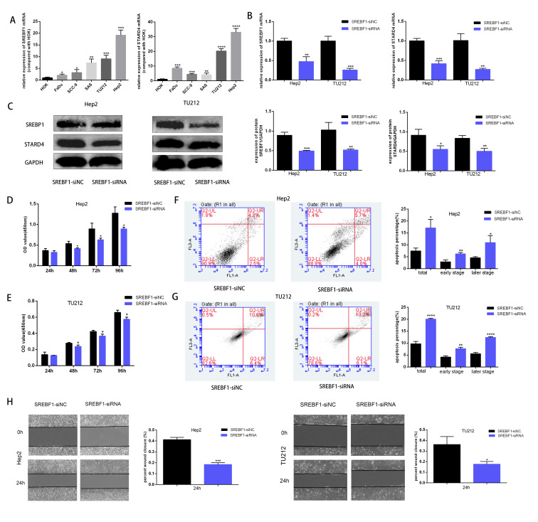
Results: SREBF1 was significantly upregulated in several tumor tissues, including HNSC, and SREBF1 overexpression was positively correlated with sample type, cancer stage, tumor grade, and lymph node stage in HNSC patients. Gene enrichment analysis revealed that SREBF1 is associated with DNA replication and homologous recombination. SREBF1 upregulation was positively correlated with the infiltration of cytotoxic cells, B cells, T cells, T helper cells, and NK CD56 bright cells in HNSC. Knockdown of SREBF1 inhibited the proliferation and migration of HNSC cells (Hep2 and TU212) and induced apoptosis by downregulating the expression of steroidogenic acute regulatory protein-related lipid transfer 4 (STARD4).
Conclusions: SREBF1 may promote HNSC proliferation, migration and inhibit apoptosis by upregulating STARD4 and affecting the level of immune cell infiltration.
Keywords: Cell proliferation; HNSC; Immune infiltration; Migration; SREBF1.
©2023 Tan et al.
Conflict of interest statement
The authors declare there are no competing interests.
Figures
Similar articles
-
Identification of ETV4 as a prognostic biomarker and correlates with immune cell infiltration in head and neck squamous cell carcinoma.Sci Rep. 2025 Feb 27;15(1):7044. doi: 10.1038/s41598-025-90731-8. Sci Rep. 2025. PMID: 40016264 Free PMC article.
-
ADORA2B promotes proliferation and migration in head and neck squamous cell carcinoma and is associated with immune infiltration.BMC Cancer. 2025 Apr 12;25(1):673. doi: 10.1186/s12885-025-14102-2. BMC Cancer. 2025. PMID: 40221657 Free PMC article.
-
LPAR2 correlated with different prognosis and immune cell infiltration in head and neck squamous cell carcinoma and kidney renal clear cell carcinoma.Hereditas. 2022 Mar 4;159(1):16. doi: 10.1186/s41065-022-00229-w. Hereditas. 2022. PMID: 35241179 Free PMC article.
-
Upregulation of HOX genes promotes cell migration and proliferation in head and neck squamous cell carcinoma.Tumour Biol. 2021;43(1):263-278. doi: 10.3233/TUB-211525. Tumour Biol. 2021. PMID: 34633333
-
CTSG restraines the proliferation and metastasis of head and neck squamous cell carcinoma by blocking the JAK2/STAT3 pathway.Cell Signal. 2025 Mar;127:111562. doi: 10.1016/j.cellsig.2024.111562. Epub 2024 Dec 11. Cell Signal. 2025. PMID: 39672353
Cited by
-
Lipid metabolism reprogramming in head and neck cancer.Front Oncol. 2023 Oct 20;13:1271505. doi: 10.3389/fonc.2023.1271505. eCollection 2023. Front Oncol. 2023. PMID: 37927468 Free PMC article. Review.
-
Sterol regulatory element binding transcription factor 1 is an important prognostic factor for colon adenocarcinoma and closely related to immune infiltration.Cytojournal. 2024 Dec 19;21:67. doi: 10.25259/Cytojournal_43_2024. eCollection 2024. Cytojournal. 2024. PMID: 39917010 Free PMC article.
-
MulNet: a scalable framework for reconstructing intra- and intercellular signaling networks from bulk and single-cell RNA-seq data.Brief Bioinform. 2025 Mar 4;26(2):bbaf081. doi: 10.1093/bib/bbaf081. Brief Bioinform. 2025. PMID: 40095604 Free PMC article.
-
SREBP1-Dependent Metabolism as a Potential Target for Breast Cancer Risk Reduction.Cancers (Basel). 2025 May 14;17(10):1664. doi: 10.3390/cancers17101664. Cancers (Basel). 2025. PMID: 40427160 Free PMC article. Review.
-
The regulatory role and mechanism of energy metabolism and immune response in head and neck cancer.Genes Dis. 2025 Mar 19;12(6):101607. doi: 10.1016/j.gendis.2025.101607. eCollection 2025 Nov. Genes Dis. 2025. PMID: 40821122 Free PMC article. Review.
References
-
- Bindea G, Mlecnik B, Tosolini M, Kirilovsky A, Waldner M, Obenauf AC, Angell H, Fredriksen T, Lafontaine L, Berger A, Bruneval P, Fridman WH, Becker C, Pages F, Speicher MR, Trajanoski Z, Galon J. Spatiotemporal dynamics of intratumoral immune cells reveal the immune landscape in human cancer. Immunity. 2013;39:782–795. doi: 10.1016/j.immuni.2013.10.003. - DOI - PubMed
-
- Binnewies M, Roberts EW, Kersten K, Chan V, Fearon DF, Merad M, Coussens LM, Gabrilovich DI, Ostrand-Rosenberg S, Hedrick CC, Vonderheide RH, Pittet MJ, Jain RK, Zou W, Howcroft TK, Woodhouse EC, Weinberg RA, Krummel MF. Understanding the tumor immune microenvironment (TIME) for effective therapy. Nature Medicine. 2018;24:541–550. doi: 10.1038/s41591-018-0014-x. - DOI - PMC - PubMed
MeSH terms
Substances
Associated data
LinkOut - more resources
Full Text Sources
Medical
Research Materials
