Non-ossifying Fibroma of Mandible in a Four-Year-Old Girl: A Case Report
- PMID: 37090356
- PMCID: PMC10117411
- DOI: 10.7759/cureus.36470
Non-ossifying Fibroma of Mandible in a Four-Year-Old Girl: A Case Report
Abstract
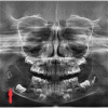
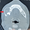
Non-ossifying fibroma (NOF) is not prevelant in the mandible. It appears mostly in the long tubular bones in children and adolescents. We are presenting a case of a four-year-old girl reported to the maxillofacial department with painless swelling over the lower right side of the jaw. On the orthopantomogram (OPG), a well-defined multilocular radiolucency with a sclerotic margin was present. On computed tomography (CT), an expansile lytic lesion with cortical thinning without a breach in cortical continuity was noted. By correlating clinical and radiological features, a diagnosis of odontogenic and/or osteogenic lesion was made. The patient was considered for an excisional biopsy with curettage. On histopathology, NOF was confirmed. On postoperative follow-up, there was no sign of recurrence, and bone regeneration was significant.
Keywords: fibrous cortical defect; histiocytic xanthogranuloma; malignant fibrous histocytoma; metaphyseal fibrous defect; non-ossifying fibroma.
Copyright © 2023, Khandaitkar et al.
Conflict of interest statement
The authors have declared that no competing interests exist.
Figures


Similar articles
-
Literature Review, Case Presentation and Management of Non-ossifying Fibroma of Right Angle of Mandible: More Than just a Cortical Defect!Indian J Otolaryngol Head Neck Surg. 2024 Feb;76(1):1054-1061. doi: 10.1007/s12070-023-04110-8. Epub 2023 Aug 7. Indian J Otolaryngol Head Neck Surg. 2024. PMID: 38440574 Free PMC article.
-
Bilateral Nonossifying Fibroma of the Mandible: A case report of a rare entity.Int J Mycobacteriol. 2023 Apr-Jun;12(2):196-206. doi: 10.4103/ijmy.ijmy_53_23. Int J Mycobacteriol. 2023. PMID: 37338484
-
Amyloid Variant of Central Odontogenic Fibroma in the Mandible: A Case Report and Literature Review.Am J Case Rep. 2020 Aug 30;21:e925165. doi: 10.12659/AJCR.925165. Am J Case Rep. 2020. PMID: 32862189 Free PMC article. Review.
-
Expansive Central Ossifying Fibroma of the Right Mandible.Cureus. 2024 Jan 24;16(1):e52863. doi: 10.7759/cureus.52863. eCollection 2024 Jan. Cureus. 2024. PMID: 38406103 Free PMC article.
-
Ossifying fibroma of the upper jaw: report of a case and review of the literature.Med Oral. 2004 Aug-Oct;9(4):333-9. Med Oral. 2004. PMID: 15292873 Review. English, Spanish.
Cited by
-
Non-ossifying fibroma of the mandible in a 4-year-old child: A rare case and review of the literature.SAGE Open Med Case Rep. 2023 Aug 6;11:2050313X231192752. doi: 10.1177/2050313X231192752. eCollection 2023. SAGE Open Med Case Rep. 2023. PMID: 37560383 Free PMC article.
-
Rare features of giant cell tumors of the bone: A case report.Exp Ther Med. 2024 Aug 28;28(5):409. doi: 10.3892/etm.2024.12698. eCollection 2024 Nov. Exp Ther Med. 2024. PMID: 39268365 Free PMC article.
-
An unusually aggressive multiple non-ossifying fibroma of the distal tibia and fibula: A case report.Bone Rep. 2023 Oct 11;19:101721. doi: 10.1016/j.bonr.2023.101721. eCollection 2023 Dec. Bone Rep. 2023. PMID: 37859796 Free PMC article.
-
Literature Review, Case Presentation and Management of Non-ossifying Fibroma of Right Angle of Mandible: More Than just a Cortical Defect!Indian J Otolaryngol Head Neck Surg. 2024 Feb;76(1):1054-1061. doi: 10.1007/s12070-023-04110-8. Epub 2023 Aug 7. Indian J Otolaryngol Head Neck Surg. 2024. PMID: 38440574 Free PMC article.
References
-
- On fibrous defects in cortical walls of growing tubular bones: their radiologic appearance, structure, prevalence, natural course, and diagnostic significance. CA J. http://pubmed.ncbi.nlm.nih.gov/14349773/ Adv Pediatr. 1955;7:13–51. - PubMed
-
- Non-ossifying fibroma of the mandible: report of a case. Park JK, Levy BA, Hanley JB Jr. http://pubmed.ncbi.nlm.nih.gov/6961119/ J Baltimore Coll Dent Surg. 1982;35:1–5. - PubMed
-
- Non-osteogenic fibroma of bone. Jaffe HL, Lichtenstein L. http://www.ncbi.nlm.nih.gov/pmc/articles/PMC2032933/ Am J Pathol. 1942;18:205–221. - PMC - PubMed
Publication types
LinkOut - more resources
Full Text Sources
