[Preprint]. 2023 May 2:2023.04.11.536376.
doi: 10.1101/2023.04.11.536376.

Optogenetic-Induced Muscle Loading Leads to Mechanical Adaptation of the Achilles Tendon Enthesis in Mice

Affiliations

Optogenetic-Induced Muscle Loading Leads to Mechanical Adaptation of the Achilles Tendon Enthesis in Mice

Elahe Ganji et al. bioRxiv. .

Update in

Abstract

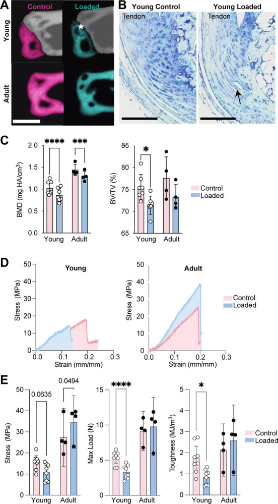
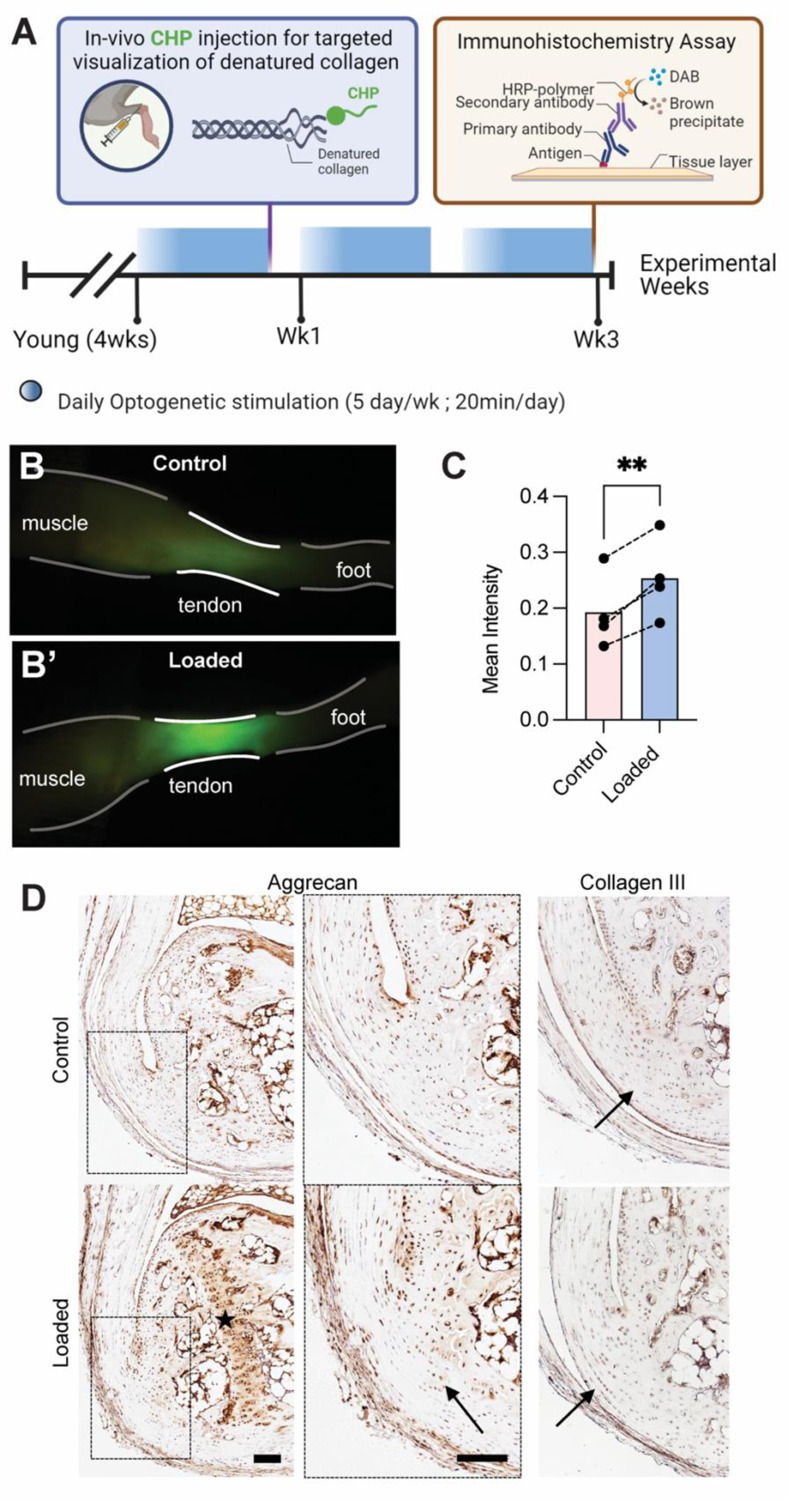
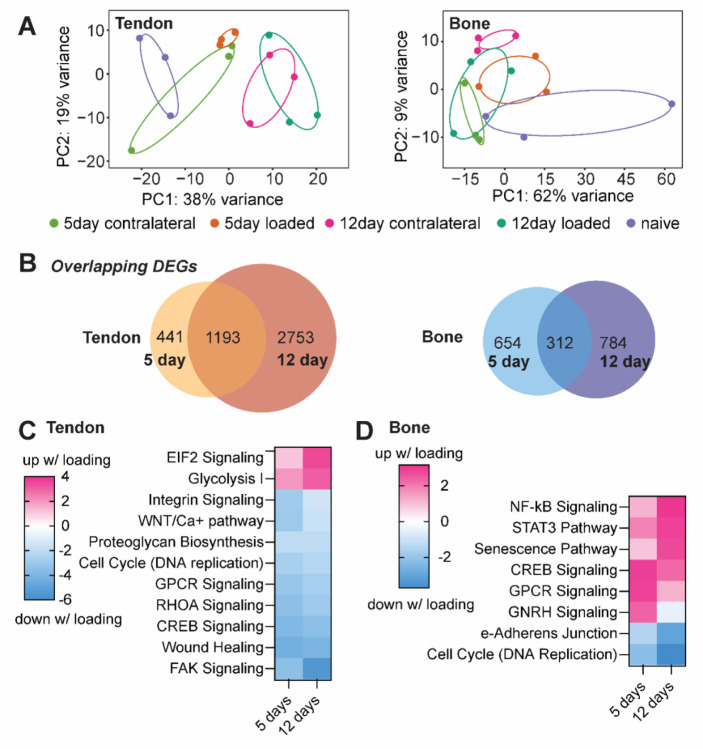
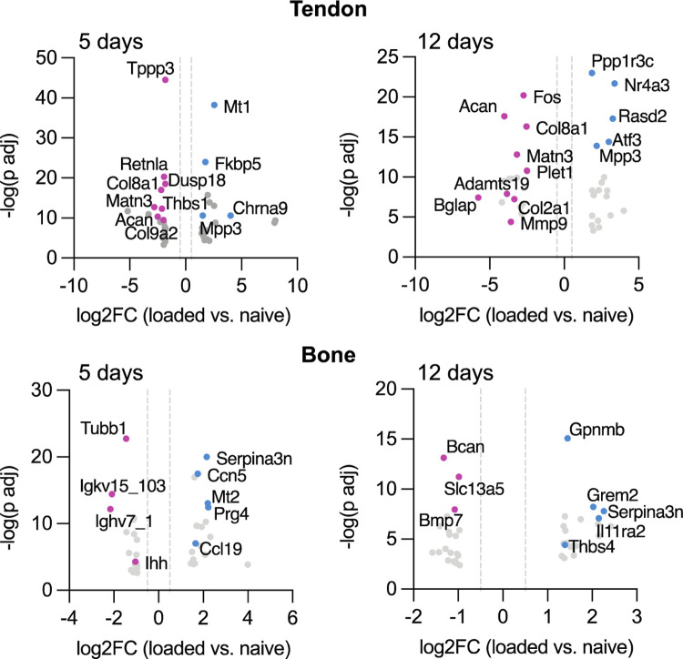
The growth of the skeleton depends on the transmission of contractile muscle forces from tendon to bone across the extracellular matrix-rich enthesis. Loss of muscle loading leads to significant impairments in enthesis development. However, little is known about how the enthesis responds to increased loading during postnatal growth. To study the cellular and matrix adaptations of the enthesis in response to increased muscle loading, we used optogenetics to induce skeletal muscle contraction and unilaterally load the Achilles tendon and enthesis in young (i.e., during growth) and adult (i.e., mature) mice. In young mice, daily bouts of unilateral optogenetic loading led to expansion of the calcaneal apophysis and growth plate, as well as increased vascularization of the normally avascular enthesis. Daily loading bouts, delivered for 3 weeks, also led to a mechanically weaker enthesis with increased molecular-level accumulation of collagen damage in young mice. However, adult mice did not exhibit impaired mechanical properties or noticeable structural adaptations to the enthesis. We then focused on the transcriptional response of the young tendon and bone following optogenetic-induced loading. After 1 or 2 weeks of loading, we identified, in tendon, transcriptional activation of canonical pathways related to glucose metabolism (glycolysis) and inhibited pathways associated with cytoskeletal remodeling (e.g., RHOA and CREB signaling). In bone, we identified activation of inflammatory signaling (e.g., NFkB and STAT3 signaling) and inhibition of ERK/MAPK and PTEN signaling. Thus, we have demonstrated the utility of optogenetic-induced skeletal muscle contraction to elicit structural, functional, and molecular adaptation of the enthesis in vivo especially during growth.


Figures

Figure 1.
Figure 1.
Optogenetic activation of triceps surae muscle group is not invasive and does not impact weight gain or generated isometric ankle joint torque in Young mice. (A) Young mice, regardless of exposure to daily bouts of optogenetic muscle stimulation (5- or 20-minute duration), gained weight for the duration of the experiment. However, adult mice lost weight from onset to end of the experiment. (B) Normalized torque significantly increased in adult but not young mice with daily bouts of 20-minute optogenetic muscle stimulation. Each data point denotes average weekly change in generated ankle torque normalized to weight of the animal. (C) The steady state generated torque at the end of the 20-minute loading bout, measured during isometric ankle plantarflexion, was ~30% of the peak force in young mice for the first week of daily optogenetic muscle loading. Error bars: Mean ± SD (*: p< 0.05, **: p< 0.001, ****: p < 0.0001, and ***: p = 0.0004).
Figure 2.
Figure 2.. Repeated loading in Young mice results in disruption of structural toughening mechanisms at enthesis.
Repeated loading resulted in (A) opening of the calcaneal growth plate, (B) disruption of enthesis tidemark, reduction of subchondral bone mineralization, and vascular infiltration, in Young enthesis and apophyses. (C) Young and Adult loaded apophyses had significantly lower BMD compared to age-matched controls. Structural adaptations in the Young enthesis and apophysis, coincides with (D, E) significantly less tensile strength and toughness in the loaded compared to non-loaded contralateral limbs. Star specifies the opening of the calcaneal growth plate. Scale bars denote (A) 500μm and (B) 100μm. (*: p< 0.0.5, **: p< 0.001, ****: p < 0.0001, and ***: p = 0.0004).
Figure 3.
Figure 3.
Repeated loading led to denatured collagen fibers and increased prevalence of ECM components at the enthesis and growth plate. (A) Schematic shows experimental design for evaluation of effect of repeated loading on ECM of the Young tendon and enthesis. (B, C) Repeated loading resulted in the accumulation of collagen denaturation in loaded tendon and enthesis. P<0.05. (D) Loaded entheses had qualitatively higher aggrecan and type III collagen present at the calcified and uncalcified fibrocartilaginous site of their attachments, respectively, compared to controls. Scale bar denotes 100 μm.
Figure 4.
Figure 4.
(A) Principal component analysis plots of differential gene expression for naïve, contralateral, and loaded tendons and bones after 5day and 12 days of loading. (B) In tendon and bone 1,193 and 312 genes, respectively, were differentially expressed after 5 and 12 days of loading. (C) In tendon, optogenetic loading induced upregulation of EIF2 signaling and Glycolysis I pathways, and downregulation of FAK and RHOA signaling pathways. (D) In bone, optogenetic loading led to activation of inflammatory pathways such as NF-B and STAT3 signaling, senescence, and CREB signaling, as well as downregulation of cell cycle (DNA replication) and epithelial adherens junction signaling.
Figure 5.
Figure 5.
After 5 days and 12 days of stimulation, daily bouts of optogenetic loading led to significant downregulation of genes associated with tendon stem cell activation and ECM synthesis in tendon and upregulation of proinflammatory markers in bone. Blue indicates genes upregulated with loading compared to naïve and pink indicates genes downregulated with loading compared to naïve. Data shown are log2 fold change (x-axis) and −log10 transformed (y-axis).

References

    1. Abraham Adam C., Shah Shivam A., Golman Mikhail, Song Lee, Li Xiaoning, Kurtaliaj Iden, Akbar Moeed, et al. 2019. “Targeting the NF-KB Signaling Pathway in Chronic Tendon Disease.” Science Translational Medicine 11 (481). 10.1126/scitranslmed.aav4319. - DOI - PMC - PubMed
    1. Achar Suraj, and Yamanaka Jarrod. 2019. “Apophysitis and Osteochondrosis: Common Causes of Pain in Growing Bones.” American Family Physician 99 (10): 610–18. - PubMed
    1. Almeida-Silveira M. I., Lambertz D., Pérot C., and Goubel F.. 2000. “Changes in Stiffness Induced by Hindlimb Suspension in Rat Achilles Tendon.” European Journal of Applied Physiology 81 (3): 252–57. 10.1007/s004210050039. - DOI - PubMed
    1. Andarawis-Puri Nelly, Sereysky Jedd B., Jepsen Karl J., and Flatow Evan L.. 2012. “The Relationships between Cyclic Fatigue Loading, Changes in Initial Mechanical Properties, and the in Vivo Temporal Mechanical Response of the Rat Patellar Tendon.” Journal of Biomechanics 45 (1): 59–65. 10.1016/j.jbiomech.2011.10.008. - DOI - PMC - PubMed
    1. Benjamin M., Toumi H., Ralphs J. R., Bydder G., Best T. M., and Milz S.. 2006. “Where Tendons and Ligaments Meet Bone: Attachment Sites (‘entheses’) in Relation to Exercise and/or Mechanical Load.” Journal of Anatomy 208 (4): 471–90. 10.1111/j.1469-7580.2006.00540.x. - DOI - PMC - PubMed

Publication types