[Preprint]. 2024 Feb 1:2023.04.12.536513.
doi: 10.1101/2023.04.12.536513.

Rbm8a deficiency causes hematopoietic defects by modulating Wnt/PCP signaling

Affiliations

Rbm8a deficiency causes hematopoietic defects by modulating Wnt/PCP signaling

Agnese Kocere et al. bioRxiv. .

Update in

  • Rbm8a deficiency causes hematopoietic defects by modulating Wnt/PCP signaling.
    Kocere A, Chiavacci E, Soneson C, Jacobson ST, Harrison EN, Méndez-Acevedo KM, MacGowan JS, Wells HH, Hiltabidle MS, Raghunath A, Shavit JA, Panáková D, Williams MLK, Robinson MD, Mosimann C, Burger A. Kocere A, et al. Dev Biol. 2025 Dec;528:34-56. doi: 10.1016/j.ydbio.2025.08.021. Epub 2025 Sep 2. Dev Biol. 2025. PMID: 40907933

Abstract

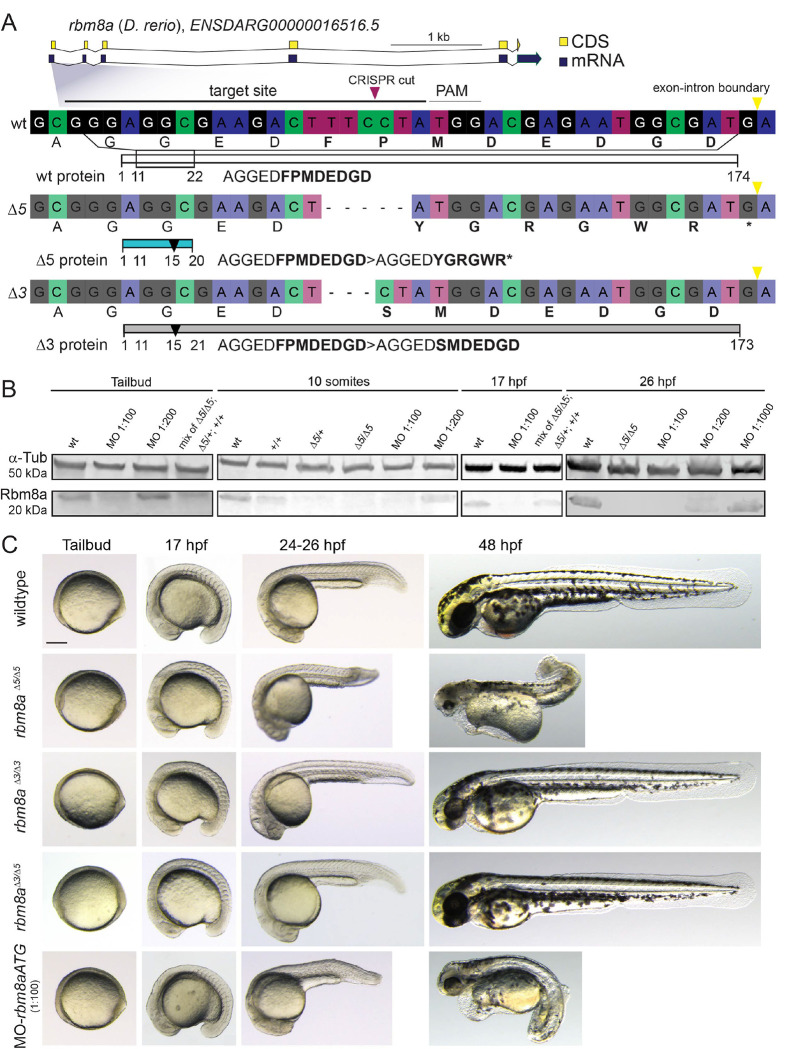
Defects in blood development frequently occur among syndromic congenital anomalies. Thrombocytopenia-Absent Radius (TAR) syndrome is a rare congenital condition with reduced platelets (hypomegakaryocytic thrombocytopenia) and forelimb anomalies, concurrent with more variable heart and kidney defects. TAR syndrome associates with hypomorphic gene function for RBM8A/Y14 that encodes a component of the exon junction complex involved in mRNA splicing, transport, and nonsense-mediated decay. How perturbing a general mRNA-processing factor causes the selective TAR Syndrome phenotypes remains unknown. Here, we connect zebrafish rbm8a perturbation to early hematopoietic defects via attenuated non-canonical Wnt/Planar Cell Polarity (PCP) signaling that controls developmental cell re-arrangements. In hypomorphic rbm8a zebrafish, we observe a significant reduction of cd41-positive thrombocytes. rbm8a-mutant zebrafish embryos accumulate mRNAs with individual retained introns, a hallmark of defective nonsense-mediated decay; affected mRNAs include transcripts for non-canonical Wnt/PCP pathway components. We establish that rbm8a-mutant embryos show convergent extension defects and that reduced rbm8a function interacts with perturbations in non-canonical Wnt/PCP pathway genes wnt5b, wnt11f2, fzd7a, and vangl2. Using live-imaging, we found reduced rbm8a function impairs the architecture of the lateral plate mesoderm (LPM) that forms hematopoietic, cardiovascular, kidney, and forelimb skeleton progenitors as affected in TAR Syndrome. Both mutants for rbm8a and for the PCP gene vangl2 feature impaired expression of early hematopoietic/endothelial genes including runx1 and the megakaryocyte regulator gfi1aa. Together, our data propose aberrant LPM patterning and hematopoietic defects as consequence of attenuated non-canonical Wnt/PCP signaling upon reduced rbm8a function. These results also link TAR Syndrome to a potential LPM origin and a developmental mechanism.

Keywords: development; hematopoiesis; morphogenesis; non-canonical Wnt; thrombocytopenia; zebrafish.


Conflict of interest statement

COMPETING INTERESTS STATEMENT J.A.S. has been a consultant for Sanofi, Takeda, Genentech, CSL Behring, and HEMA Biologics.

Figures

Figure 1:
Figure 1:. Genetic perturbations in the zebrafish rbm8a gene.
(A) Allelic series of rbm8a in zebrafish with wild type allele, rbm8aΔ5 allele and rbm8aΔ3 allele induced by Cas9-mediated mutagenesis. (B) Western blot analysis of Rbm8a protein in mutants and morphants at different developmental stages, showing gradual decrease of maternal protein in mutants and translation block in morphants. (C) Representative images of zebrafish embryos carrying different rbm8a allele combinations during early development. Compared to wild type and different rbm8aΔ5 and rbm8aΔ3 combinations, homozygous rbm8aΔ5/Δ5 carriers display severe microcephaly and corkscrew tail phenotype. rbm8a morphant (1:100 morpholino dilution) phenotypically recapitulates the homozygous rbm8aΔ5/Δ5 embryos. Scale bars in (C): 250 μm, applies to all panels in C.
Figure 2:
Figure 2:. rbm8a perturbation reduces thrombocyte numbers in zebrafish larvae.
(A) Workflow schematic of cd41:EGFP-positive thrombocyte progenitor quantification from circulation and from the caudal hematopoietic territory (CHT). Created with Biorender.com (Subscription: Individual, Agreement number: KV25WQCJKF). (B-I) Representative fluorescent dissecting scope and confocal images of zebrafish embryos transgenic for the thrombocyte marker cd41:EGFP, greyscale and color-inverted to reveal GFP-positive cells; anterior to the left, insert depicts brightfield image of larvae for reference. (J,K) cd41:EGFP-positive thrombocyte counts at 3 dpf (J) and 6 dpf (K) for each analyzed condition. Wild type larvae have significantly more cd41:EGFP-positive cells at 3 and 6 days compared to the high morpholino dose (1:125 and 1:150, only at 3 dpf), while lower dose (1:200) remained at wild type levels (J,K). At 6 dpf (K), viable trans-heterozygous allele combinations for rbm8a also show a significant thrombocyte reduction. Note that morpholino injections will block translation from maternal mRNA, while mutant combinations will retain maternal mRNA function from the wild type allele carried by the mother. Individual datapoints (total number of cd41:GFP-positive cells per embryo) shown with mean and standard deviation, significance calculated by Mann-Whitney test: 3 dpf (J) wild type vs. control MO p=0.7756 (not significant), wild type vs. MO-rbm8aATG 1:125 p<0.0001, wild type vs. MO-rbm8aATG 1:150 p<0.0001, wild type vs. MO-rbm8aATG 1:175 p=0.2589 (not significant), wild type vs. MO-rbm8aATG 1:200 p=0.5628 (not significant), rbm8a+/+ vs. rbm8aΔ5/+ p=0.605 (not significant), rbm8a+/+ vs. rbm8aΔ3/ Δ3 p=0.7655 (not significant), rbm8a+/+ vs. rbm8aΔ3/ Δ5 p=0.4289 (not significant); 6 dpf (K) wild type vs. control MO p=0.9567 (not significant), wild type vs. MO-rbm8aATG 1:125 p<0.0001, wild type vs. MO-rbm8aATG 1:150 p=0.5198 (not significant), wild type vs. MO-rbm8aATG 1:175 p=0.7147 (not significant), wild type vs. MO-rbm8aATG 1:200 p=0.288 (not significant), rbm8a+/+ vs. rbm8aΔ5/+ p=0.9462 (not significant), rbm8a+/+ vs. rbm8aΔ3/ Δ3 p=0.0208, rbm8a+/+ vs. rbm8aΔ3/ Δ5 p<0.0001 (see Supplementary Data 2 for details). Scale bars in (B): 200 μm, applies to panels B,D,F,H; (C): 150 μm, applies to panels C,E,G,I.
Figure 3:
Figure 3:. Intron retention in mRNAs encoding non-canonical Wnt/PCP components in zebrafish rbm8a mutants.
(A) Experimental design of bulk RNA-seq experiment. Created with Biorender.com (Subscription: Individual, Agreement number: SN25WQAKB9). (B,C) Volcano plots of tailbud (B) and 24 hpf (C) comparisons between wild type versus rbm8a-mutant embryos; significantly down- (magenta) and up-regulated (blue) genes with named examples (see Supplementary Data 3 and 4 for details). (D) Qualitative assessment of retained introns between wild type versus rbm8a-mutant embryos; note that retained introns are already apparent at tailbud stage (see Supplementary Data 6 for details). (E, F) Read coverage plot of mRNA sequencing reads of non-canonical Wnt/PCP components celsr2, wnt11f2 (former wnt11), and vangl2 that feature mRNAs with retained introns (E,F) and/or differential transcript usage (G) in rbm8a-mutant embryos. (H) Differential transcript usage of vangl2 transcripts at 24 hpf (exon 1 followed by long intron, asterisks).
Figure 4:
Figure 4:. rbm8a and vangl2 perturbations reduce the rate of posterior LPM growth.
(A) Schematic of the workflow to image the zebrafish using Light sheet microscope from 4 different angles, assemble in a 3D image (represented by the globe) and measure volume and area of the PLPM scl:GFP surface rendering (represented by the map). Created with Biorender.com (Subscription: Individual, Agreement number: IG25WQA6FD). (B-D) Surface rendering of GFP signal from light sheet-based timelapse imaging of scl:GFP transgenic zebrafish embryos. Wild type (A), rbm8a morphant (B), and vangl2 morphant (C) embryos at 5, 7, 9 and 11 somite stages, dorsal view, anterior to the top, posterior end of the LPM at bottom. (D,E) Area and volume of scl:GFP-expressing LPM territory measured with Imaris, comparing wild type (n=4), rbm8a morphant (n=5) and vangl2 morphant (n=4) measurements depicted in color groups corresponding to sample groups. Note how area in wild type increases by x^2 (D) and volume by x^3 (E), revealing reduced growth rates of the posterior scl:GFP territory upon rbm8a and vangl2 perturbations. Datapoints shown are average with standard deviation, significance calculated by 2-way Anova. Area (p<0.0001) and volume (p<0.0001) are significantly different in MO-rbm8aATG- and MO1-vangl2-injected embryos compared to stage-matched wild type embryos at the analyzed timepoints (see Supplementary Data 9 for details). Scale bar in (B): 150 μm, applies to all panels in B-D.
Figure 5:
Figure 5:. rbm8a perturbation results in convergence and extension defects.
(A) mRNA in situ hybridization for myoD, dlx3b, tbxta, and hgg1 as landmarks to measure morphometric parameters associated with convergence and extension as influenced by non-canonical Wnt/PCP signaling. Zebrafish embryos are shown as lateral views (odd columns, anterior to the top, ventral to the left) and dorsal views (even columns, anterior to the top). Black arrowheads depict anterior and posterior extent of the body axis in lateral views, pink and grey arrowheads depict otic placodes and somites at measured position in dorsal views. Axis length and axis angle (marked by black arrowheads) were measured from the odd panels. Neural plate (between the otic placodes at beginning of notochord, pink arrowheads) and somite width (last 3 somites, grey arrowheads) were measured from the even panels. (B-E) Morphometric measurements for axis length (B), axis angle (C), neural plate width (D), and somite width (E) from individual embryo (wild type, mutant, morphant) images at different developmental stages. Datapoints shown are average with standard deviation, significance calculated by Mann Whitney test: axis length (B) 6–7 somite stage wild type vs. MO-rbm8aATG p<0.0001, wild type vs. rbm8aΔ5/Δ5 p<0.0001, wild type vs. rbm8aΔ3/Δ5 p=0.4394 (not significant), wild type vs. rbm8aΔ3/Δ3 p=0.9347 (not significant), 8–9 somite stage wild type vs. MO-rbm8aATG p<0.0001, wild type vs. rbm8aΔ5/Δ5 p<0.0001, wild type vs. rbm8aΔ3/Δ5 p=0.0048, wild type vs. rbm8aΔ3/Δ3 p=0.1643 (not significant), 10–11 somite stage wild type vs. MO-rbm8aATG p=0.0001, wild type vs. rbm8aΔ5/Δ5 p=0.0003, wild type vs. rbm8aΔ3/Δ5 p=0.0945 (not significant), wild type vs. rbm8aΔ3/Δ3 p=0.1219 (not significant); axis angle (C) 6–7 somite stage wild type vs. MO-rbm8aATG p<0.0001, wild type vs. rbm8aΔ5/Δ5 p<0.0001, wild type vs. rbm8aΔ3/Δ5 p=0.3325 (not significant), wild type vs. rbm8aΔ3/Δ3 p=0.4449 (not significant), 8–9 somite stage wild type vs. MO-rbm8aATG p<0.0001, wild type vs. rbm8aΔ5/Δ5 p<0.0001, wild type vs. rbm8aΔ3/Δ5 p=0.7492 (not significant), wild type vs. rbm8aΔ3/Δ3 p=0.2633 (not significant), 10–11 somite stage wild type vs. MO-rbm8aATG p<0.0001, wild type vs. rbm8aΔ5/Δ5 p<0.0001, wild type vs. rbm8aΔ3/Δ5 p=0.439 (not significant), wild type vs. rbm8aΔ3/Δ3 p=0.342 (not significant), neural plate width (D) 6–7 somite stage wild type vs. MO-rbm8aATG p=9978 (not significant), wild type vs. rbm8aΔ5/Δ5 p<0.0001, wild type vs. rbm8aΔ3/Δ5 p=0.4437 (not significant), wild type vs. rbm8aΔ3/Δ3 p=0.3001 (not significant), 8–9 somite stage wild type vs. MO-rbm8aATG p=0.0086, wild type vs. rbm8aΔ5/Δ5 p<0.0001, wild type vs. rbm8aΔ3/Δ5 p<0.0001, wild type vs. rbm8aΔ3/Δ3 p=0.0008, 10–11 somite stage wild type vs. MO-rbm8aATG p=0.8324 (not significant), wild type vs. rbm8aΔ5/Δ5 p=0.0198, wild type vs. rbm8aΔ3/Δ5 p=0.5477 (not significant), wild type vs. rbm8aΔ3/Δ3 p=0.5448 (not significant); somite width (E) 6–7 somite stage wild type vs. MO-rbm8aATG p<0.0001, wild type vs. rbm8aΔ5/Δ5 p<0.0001, wild type vs. rbm8aΔ3/Δ5 p=0.2296 (not significant), wild type vs. rbm8aΔ3/Δ3 p=0.7951 (not significant), 8–9 somite stage wild type vs. MO-rbm8aATG p=0.1853 (not significant), wild type vs. rbm8aΔ5/Δ5 p<0.0001, wild type vs. rbm8aΔ3/Δ5 p=0.2642 (not significant), wild type vs. rbm8aΔ3/Δ3 p=0.6899 (not significant), 10–11 somite stage wild type vs. MO-rbm8aATG p=0.0028, wild type vs. rbm8aΔ5/Δ5 p=0.0003, wild type vs. rbm8aΔ3/Δ5 p=0.5769 (not significant), wild type vs. rbm8aΔ3/Δ3 p=0.4317 (not significant) (see Supplementary Data 10 for details). Scale bar in (A): 250 μm, applies to all panels in A.
Figure 6:
Figure 6:. rbm8a attenuation sensitizes zebrafish to non-canonical Wnt/PCP defects.
(A-F) 2 dpf lateral views of representative zebrafish larvae analyzed for each genetic perturbation. Anterior to the left. (A) Uninjected wild type as phenotype reference. (B) Suboptimal (1:500) MO-rbm8aATG injection that does not result in a visible phenotype (as compared to higher doses) and provides a baseline for rbm8a attenuation. (C-F) Representative phenotypes following sequential injection of suboptimal MO-rbm8aATG and suboptimal doses of individual, validated morpholinos against the non-canonical Wnt/PCP components fzd7 (C), vangl2 (D), wnt5b (E), and wnt11f2 (F). (G) Observed phenotypes in percent of embryos. (H-K) Reduced function of vangl2 and wnt5b results in reduced cd41:GFP-expressing thrombocytes at 6 dpf. (H-J) Lateral, inverted greyscale views of 6 dpf cd41:GFP-transgenic embryos confocal-imaged for GFP fluorescence, inserts depict brightfield view of imaged embryos and overall phenotype reference. Note how suboptimal dosing retains longer viability and less morphological impact (compare to D,E). (K) Thrombocyte quantification at 6 dpf reveals mild, yet dose-dependent reduction in cell number upon vangl2 and wnt5b perturbation. Individual datapoints (total number of cd41:GFP- positive cells per embryo) shown with mean and standard deviation, significance calculated by Mann-Whitney test: 6 dpf (K) wild type vs. control MO p=0.4981 (not significant), wild type vs. MO2-wnt5b 1:10 p=0.0407, wild type vs. MO2-wnt5b 1:15 p=0.2364 (not significant), wild type vs. MO1-vangl2 1:5 p=0.0533 (not significant), wild type vs. MO1-vangl2 1:10 p=0.3828 (not significant) (see Supplementary Data for 11 for details). Scale bar in (A): 100 μm, applies to panels A-F; (H): 150 μm, applies to panels H-J.
Figure 7:
Figure 7:. Select changes in hematopoietic marker expression in rbm8a- and vangl2-mutant zebrafish embryos.
(A) Schematic of basic hematopoietic lineage and marker relationships. Created with Biorender.com (Subscription: Individual, Agreement number: IZ25WQERBS). AGM: aorta-gonadal-mesonephros area; ICM: intermediate cell mass; PBI: posterior blood island; CHT: caudal hematopoietic territory; VDA: ventral dorsal aorta. (B-F) Expression patterns for individual marker genes in wild type, morpholino-injected, rbm8a- and vangl2-mutant zebrafish embryos at stage-matched 18 somites. Schematic of zebrafish embryo in the last panel in B-H shows where marker expression is expected and analyzed. (B) myoD as marker for somitic muscles shows comparable expression across conditions, with vangl2 mutants depicting the short, deteriorating tail typical for these mutants (asterisk). (C) The early endothelial marker sox7 is reduced in the trunk upon rbm8a perturbation (arrowheads) and seemingly normal in vangl2 mutants (asterisk). (D) The endothelial marker kdrl is decreased similarly as sox7. (E) The erythroid marker gata1 is reduced in all rbm8a-perturbed embryo conditions (arrowheads) and only retained in posterior-most LPM cells of vangl2 mutants (asterisk). (F) The hematopoietic progenitor marker gfi1aa is reduced in all rbm8a-perturbed embryo conditions (arrowheads) and barely detectable in vangl2-mutant embryos (asterisk). (G) In contrast, gfi1b is unchanged in all conditions. (H) The hematopoietic progenitor marker runx1 in rbm8a morphants and mutants is reduced or absent in the trunk and posterior blood island (PBI) (embryo midline, future dorsal aorta, arrowheads); vangl2 mutants also show reduced trunk expression (arrowhead) yet retain PBI runx1 expression (asterisk). (I) mRNA in situ hybridization-based quantification n of gata1, gfi1aa, and gfi1b expression levels; individual data points (normalized signal intensity/embryo) shown with mean and standard deviation, significance calculated by Mann-Whitney test: gata1 ISH signal intensity in wild type vs. MO-rbm8aATG p<0.0001, gfi1aa ISH signal intensity in wild type vs. MO-rbm8aATG p=0,031, gfi1b ISH signal intensity in wild type vs. MO-rbm8aATG p=0.7727 (not significant). (J-M) mRNA in situ hybridization-based quantification of runx1 in select areas (aorta-gonadal-mesonephros area (AGM): embryo midline, future dorsal aorta), posterior blood island (PBI), neurons). (J) Representative scale of runx1 signal ranked from low to high in rbm8a wild type siblings. The numbers on the bottom represent the average scores for AGM, PBI and neurons, respectively. (K) Signal in AGM. (L) Signal in PBI. (M) Signal in dorsal neurons. Individual data points (average of 4 scores per embryo) shown with mean and standard deviation, significance calculated by Mann-Whitney test: runx1a signal intensity in AGM (K) in wild type vs. rbm8aΔ5/+ p=0.0048, wild type vs. rbm8aΔ5/Δ5 p<0.0001, rbm8aΔ5/+ vs. rbm8aΔ5/Δ5 p=0.0009; PBI (L) in wild type vs. rbm8aΔ5/+ p=0.0077, wild type vs. rbm8aΔ5/Δ5 p<0.0001, rbm8aΔ5/+ vs. rbm8aΔ5/Δ5 p=0.0007; neurons (M) wild type vs. rbm8aΔ5/+ p=0.1604 (not significant), wild type vs. rbm8aΔ5/Δ5 p=0.005, rbm8aΔ5/+ vs. rbm8aΔ5/Δ5 p=0.0481. Violin plot in background depicts population of the not averaged scores. Numbers (n)=average/total (see Supplementary Data 12 for details). Scale bar in (B): 100 μm, applies to all panels in B-H.

References

    1. Petit F. & Boussion S. Thrombocytopenia Absent Radius Syndrome. GeneReviews® (2022). - PubMed
    1. Albers C. A., Newbury-Ecob R., Ouwehand W. H. & Ghevaert C. New insights into the genetic basis of TAR (thrombocytopenia-absent radii) syndrome. Curr Opin Genet Dev 23, 316–23 (2013). - PubMed
    1. Klopocki E. et al. Complex inheritance pattern resembling autosomal recessive inheritance involving a microdeletion in thrombocytopenia-absent radius syndrome. Am J Hum Genet 80, 232–240 (2007). - PMC - PubMed
    1. Greenhalgh K. L. Thrombocytopenia-absent radius syndrome: a clinical genetic study. J Med Genet 39, 876–881 (2002). - PMC - PubMed
    1. Boussion S. et al. TAR syndrome: clinical and molecular characterization of a cohort of 26 patients and description of novel non-coding variants of RBM8A. Hum Mutat 41, 1220–1225 (2020). - PubMed

Publication types