[Preprint]. 2023 Mar 13:2023.03.10.532018.
doi: 10.1101/2023.03.10.532018.

Loss of MC1R signaling implicates TBX3 in pheomelanogenesis and melanoma predisposition

Affiliations

Loss of MC1R signaling implicates TBX3 in pheomelanogenesis and melanoma predisposition

H Matthew Berns et al. bioRxiv. .

Abstract

The human Red Hair Color (RHC) trait is caused by increased pheomelanin (red-yellow) and reduced eumelanin (black-brown) pigment in skin and hair due to diminished melanocortin 1 receptor (MC1R) function. In addition, individuals harboring the RHC trait are predisposed to melanoma development. While MC1R variants have been established as causative of RHC and are a well-defined risk factor for melanoma, it remains unclear mechanistically why decreased MC1R signaling alters pigmentation and increases melanoma susceptibility. Here, we use single-cell RNA-sequencing (scRNA-seq) of melanocytes isolated from RHC mouse models to reveal a Pheomelanin Gene Signature (PGS) comprising genes implicated in melanogenesis and oncogenic transformation. We show that TBX3, a well-known anti-senescence transcription factor implicated in melanoma progression, is part of the PGS and binds both E-box and T-box elements to regulate genes associated with melanogenesis and senescence bypass. Our results provide key insights into mechanisms by which MC1R signaling regulates pigmentation and how individuals with the RHC phenotype are predisposed to melanoma.

Keywords: TBX3; melanocortin 1 receptor (MC1R); melanocytes; melanoma; red hair color.


Conflict of interest statement

Competing Interest Statement: Authors have no competing interests to declare.

Figures
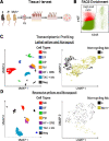
Figure 1.
Figure 1.. Overview of single cell sequencing of primary murine melanocytes.
(A) Schematic of tissue harvest and digestion for melanocyte isolation from dorsal skin (a = nonagouti; Ay = lethal yellow, Mc1re = recessive yellow). (B) Representative FACS enrichment of c-KIT+/CD45 cells isolated from the lethal yellow sample (red). (C-D) Uniform Manifold Approximation and Projection (UMAP) plots of the sequenced cells that passed quality control for each pairwise experiment. Among the cells isolated from nonagouti and lethal yellow paired samples (6,413 cells in C) and nonagouti and recessive yellow paired samples (4645 cells in D), skin cell types were identified and labeled as follows: Melanocytes (Mc), Dermal fibroblasts (DF), Dermal papilla (DP), Epidermis (Epi), Hair follicle and Outer root sheath (HF + ORS), and Transit amplifying and Matrix cells (TAC + Mx). Right panels show UMAP plots of the 2256 cells classified as non-cycling melanocytes. The nonagouti melanocytes (black) segregate from the recessive yellow and lethal yellow (gold) melanocytes consistent with a eumelanin and pheomelanin producing melanocytes having distinct transcriptional profiles.
Figure 2.
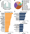
Figure 2.. Enriched processes and pathways present in the PGS.
(A) Top 15 significant GO terms ranked by fold-enrichment (FDR < 0.05). (B) Top 15 significant pathway terms from IPA ranked by significance (p-value <0.05).
Figure 3.
Figure 3.. Identifying PGS regulatory factors.
(A) List of all differentially expressed transcription/co-transcription factors found within the PGS and ranked by FDR from the nonagouti/lethal yellow pairwise differential analysis. (B) Top 10 predicted activated (green) and inhibited (red) regulators of the PGS gene list ranked by Z-score.
Figure 4.
Figure 4.. ChIP-seq analysis implicates TBX3 as a regulator of melanogenesis.
(A) Position of called TBX3 peaks relative to the TSS. (B) ChIPseeker plot identifying genomic regions in which TBX3 is bound. (C) Top 15 GO terms of genes associated with TBX3 ChIP target genes ranked by −Log10 of the FDR. (D) Top 5 predicted phenotype associations (human and mouse) from TBX3 ChIP gene by −Log10 of the FDR.
Figure 5.
Figure 5.. TBX3 binds MITF targets at E-boxes.
(A) The top 10 motifs enriched within TBX3 ChIP peaks show over-representation of core E-box (red) and T-box (purple) sequences (B) The majority of TBX3 ChIP peaks contain either a T-box motif (14%), E-box motif (27%), or both (32%). (C) Top motifs of shared MITF and TBX3 peak sites (E-box = red; T-box = purple). (D) CentriMo plot showing top 9 centrally bound motifs.
Figure 6.
Figure 6.. TBX3 binds the E-box of pigmentation gene targets.
(A) Intersection showing the number of murine genes (4072) associated with ChIP peaks in both our TBX3 dataset and a published MITF dataset. (B) Intersection of PGS genes identified as targets of both TBX3 and MITF. (C) UCSC browser view of TBX3 and MITF ChIP peaks associated with pigmentation genes. Browser tracks are as follows: TBX3 = replicated binding sites in our dataset; MITF = ChIP peaks from Strub et al; cCRE=encode annotated candidate cis-regulatory elements. The location of verified MITF regulatory elements containing E-box motifs are indicated (*) (refs in text).
Figure 7.
Figure 7.. TBX3 binds to and regulates PGS genes.
(A) Heatmap depicting relative gene expression of 2780 DEG (Adjusted p-value < 0.05) from the TBX3 knockdown samples against a non-coding control. (B) Top KEGG terms ranked by −Log10 of the p-value for upregulated (green) and downregulated (red) genes. (C) Intersection of DE TBX3 gene targets against PGS TBX3 targets.

References

    1. Ha L., Merlino G. & Sviderskaya E. V. Melanomagenesis: Overcoming the barrier of melanocyte senescence. Cell Cycle 7, 1944–1948 (2008). - PMC - PubMed
    1. Bennett D. C. & Medrano E. E. Molecular Regulation of Melanocyte Senescence. Pigm Cell Res 15, 242–250 (2002). - PubMed
    1. Vredeveld L. C. W. et al. Abrogation of BRAFV600E-induced senescence by PI3K pathway activation contributes to melanomagenesis. Gene Dev 26, 1055–1069 (2012). - PMC - PubMed
    1. Wagstaff W. et al. Melanoma --- Molecular genetics, metastasis, targeted therapies, immunotherapies, and therapeutic resistance. Genes Dis 9, 1608–1623 (2022). - PMC - PubMed
    1. Rhodes A. R., Weinstock M. A., Fitzpatrick T. B., Mihm M. C. & Sober A. J. Risk Factors for Cutaneous Melanoma: A Practical Method of Recognizing Predisposed Individuals. Jama 258, 3146–3154 (1987). - PubMed

Publication types