[Preprint]. 2023 Apr 13:rs.3.rs-2721176.
doi: 10.21203/rs.3.rs-2721176/v1.

SMYD2 Regulates Vascular Smooth Muscle Cell Phenotypic Switching and Intimal Hyperplasia via Interaction with Myocardin

Affiliations

SMYD2 Regulates Vascular Smooth Muscle Cell Phenotypic Switching and Intimal Hyperplasia via Interaction with Myocardin

Yu Zhou et al. Res Sq. .

Update in

Abstract

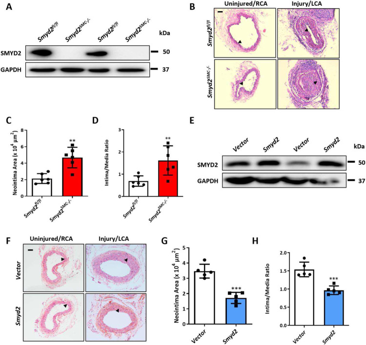
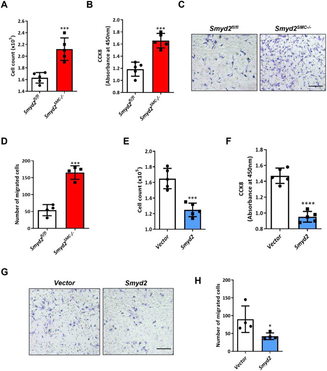
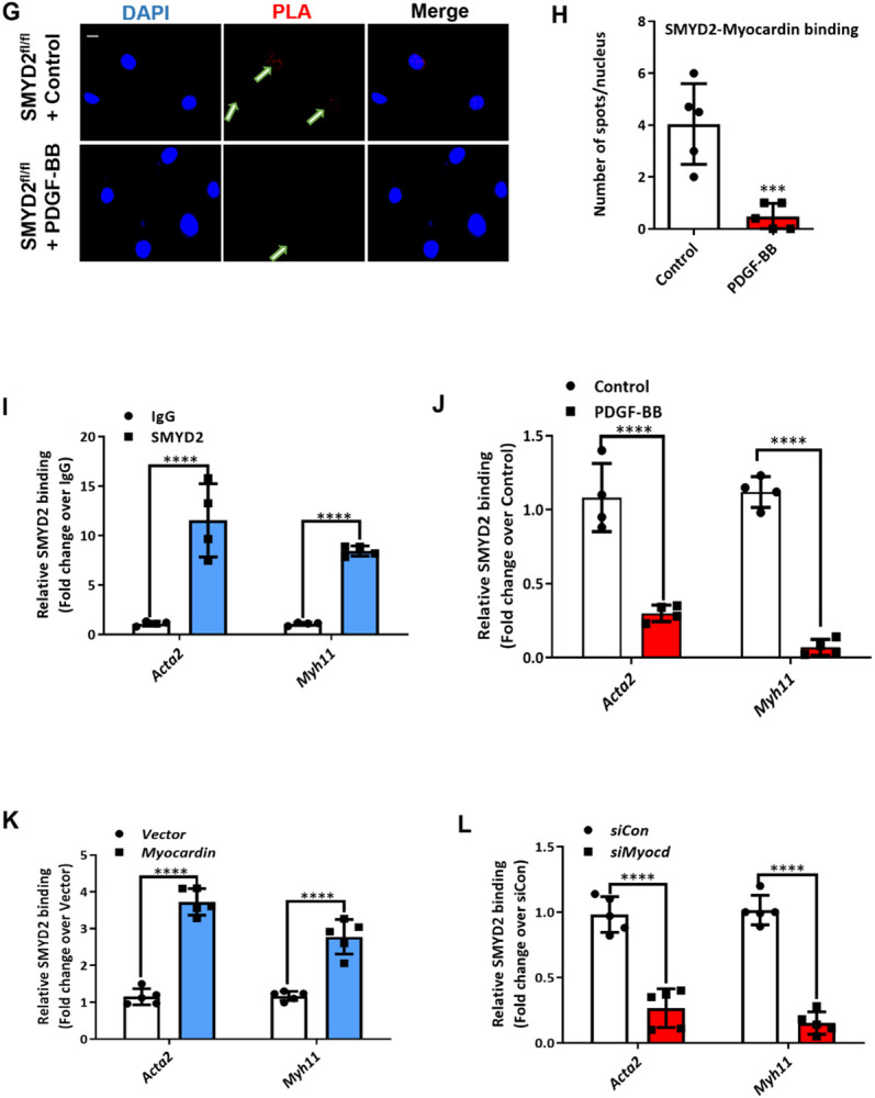
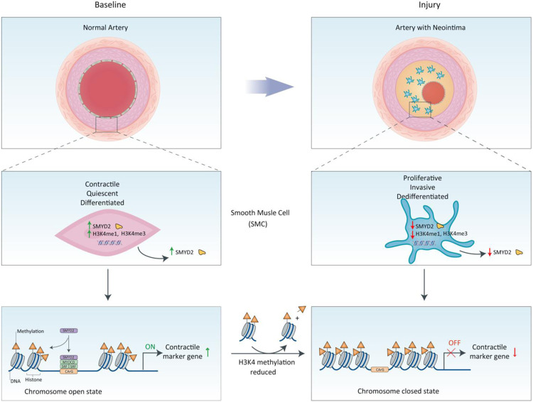
The SET and MYND domain-containing protein 2 (SMYD2) is a histone lysine methyltransferase that has been reported to regulate carcinogenesis and inflammation. However, its role in vascular smooth muscle cell (VSMC) homeostasis and vascular diseases has not been determined. Here, we investigated the role of SMYD2 in VSMC phenotypic modulation and vascular intimal hyperplasia and elucidated the underlying mechanism. We observed that SMYD2 expression was downregulated in injured carotid arteries in mice and phenotypically modulated VSMCs in vitro. Using a SMC-specific Smyd2 knockout mouse model, we found that Smyd2 ablation in VSMCs exacerbates neointima formation after vascular injury in vivo. Conversely, Smyd2 overexpression inhibits VSMC proliferation and migration in vitro and attenuates arterial narrowing in injured vessels in mice. Smyd2 downregulation promotes VSMC phenotypic switching accompanied with enhanced proliferation and migration. Mechanistically, genome-wide transcriptome analysis and loss/gain-of-function studies revealed that SMYD2 up-regulates VSMC contractile gene expression and suppresses VSMC proliferation and migration, in part, by promoting expression and transactivation of the master transcription cofactor myocardin. In addition, myocardin directly interacts with SMYD2, thereby facilitating SMYD2 recruitment to the CArG regions of SMC contractile gene promoters and leading to an open chromatin status around SMC contractile gene promoters via SMYD2-mediated H3K4 methylation. Hence, we conclude that SMYD2 is a novel regulator of VSMC contractile phenotype and intimal hyperplasia via a myocardin-dependent epigenetic regulatory mechanism and may be a potential therapeutic target for occlusive vascular diseases.


Conflict of interest statement

Conflict of interest

All authors declare no potential conflicts of interest.

Figures

Figure 1.
Figure 1.. SMYD2 expression was down-regulated in injured mouse carotids and phenotypically modulated VSMCs in vitro.
A, SMYD2 expression in carotid arteries of C57BL/6 mice following ligation injury of left common carotid arteries (LCA) for 7, 14 and 28 days. The uninjured right carotid arteries (RCA) were used as controls. B, Densitometric analysis of SMYD2 expression in mouse carotid arteries normalized to GAPDH (n=3 independent experiments). C, Expression of Smyd2 and SM marker genes (Acta2 and Cnn1) was determined by RT-qPCR in different passages (P0 and P6) of primary mouse aortic VSMCs cultured in 10% FBS + DMEM, values were normalized to β-actin (n=5 independent experiments). D, Western blot analysis of SMYD2 and SM22α in human VSMCs (HASMCs) upon treatment with PDGF-BB (20 ng/ml) for various durations (0-48 h), followed by serum starvation for 24 h. GAPDH served as the loading control. For all bar graphs, data are expressed as the means ± SEM, *P < 0.05 (unpaired, two-tailed Student’s t-test).
Figure 2.
Figure 2.. SMC-SMYD2 regulated neointima formation in the carotid arteries after injury in mice.
A, Western blot analysis of SMYD2 protein in primary aortic VSMCs isolated from Smyd2fl/fl control mice and Smyd2SMC−/− mice. B, Representative images of H&E-stained cross-sections of carotid arteries from Smyd2fl/fl control mice and Smyd2SMC−/− mice following sham or ligation injury for 14 days. Arrows indicated internal elastic lamina (IEL), Scale bar: 100 μm. C and D, Quantitative analysis of neointima area (C) and intima-to-media ratio (D) at day 14 post-injury (n=6 mice per group). E, GFP vector or GFP-SMYD2 lentiviruses (1×10⁹) were injected immediately around sham-operated or injured carotid arteries in C57BL/6 mice. Expression of SMYD2 and GAPDH in carotid arteries of GFP vector or GFP-SMYD2 group 3 days post-injury/injection was examined by Western blotting. F, Representative images of H&E-stained sections of carotid arteries from C57BL/6 mice locally injected with GFP vector control or GFP-SMYD2 overexpression lentivirus, at day 21 post-injury. Arrows indicated IEL. Scale bar: 100 μm. G and H, Quantitative analysis of neointima area (G) and intima-to-media ratio (H) at day 21 post-injury (n=5 mice per group). For all bar graphs, data are expressed as the means ± SEM, **P < 0.01, ***P < 0.001 and ****P < 0.0001 (unpaired, two-tailed Student’s t-test).
Figure 3.
Figure 3.. SMYD2 inhibited VSMC proliferation and migration in vitro.
A and B, Primary aortic VSMCs isolated from Smyd2fl/fl or Smyd2SMC−/− mice were serum-starved for 24 h, and then treated with PDGF-BB (10 ng/mL) for 24 h for the cell counting analysis (A) and CCK8 proliferation assay (B) (n=5 independent experiments). C, Transwell migration assay of murine aortic VSMCs from Smyd2fl/fl or Smyd2SMC−/− mice upon stimulation with PDGF-BB for 16 h. Scale bar, 100 μm. D, Quantification of migrated murine VSMCs (n=4 independent experiments). E and F, Primary C57BL/6 mouse aortic VSMCs were transduced with lentivirus encoding GFP-SMYD2 or GFP vector control. Post-transduction 48 h, cells were serum starved for 24 h, followed by PDGF-BB treatment. Proliferation of the transduced VSMCs was determined by cell counting analysis (E) and CCK8 proliferation assay (F) upon treatment with PDGF-BB for 24 h (n=5 independent experiments). G, Transwell migration assay of C57BL/6 mouse aortic VSMCs upon stimulation with PDGF-BB for 16 h. Scale bar, 200 μm. H, Quantification of migrated murine VSMCs. (n=4 independent experiments). For all bar graphs, data are expressed as the means ± SEM, *P < 0.05, ***P < 0.001 and ****P < 0.0001 vs. Smyd2fl/fl or vector group (unpaired, two-tailed Student’s t-test).
Figure 4.
Figure 4.. SMYD2 promoted VSMC marker expression and modulated VSMC phenotypes
A, Hierarchical clustering heat map of mRNAs differentially expressed between primary aortic VSMCs isolated from Smyd2fl/fl control mice and Smyd2SMC−/− mice, with cutoff of FC (fold change) ≥2 and adjusted P value ≤0.001; n=3 each group. B, Volcano plot, with light blue dots representing significantly downregulated protein-coding genes (n=914) and red dots representing significantly upregulated protein-coding genes (n=1176) in primary aortic VSMCs of Smyd2SMC−/− mice (q Value = adj P value). C, Gene ontology (GO) analysis of biological functions related with the differentially expressed genes in Smyd2-deficient murine VSMCs. Significant GO terms enriched in differentiated genes were plotted based on P values. D, Heat map of significantly downregulated contractile genes in Smyd2-null VSMCs compared with control VSMCs; (n=3 each group). RT-qPCR analysis (n=5 independent experiments) (E) and Western blot analysis (F) of SMC markers and myocardin in primary aortic VSMCs isolated from Smyd2fl/fl or Smyd2SMC−/− mice. RT-qPCR analysis (n=5 independent experiments) (G) and Western blot analysis (H) of SMC contractile proteins in primary VSMCs transduced with GFP or GFP-Smyd2 lentivirus. For all bar graphs, data are expressed as the means ± SEM, ***P < 0.001 and ****P < 0.0001 (unpaired, two-tailed Student’s t-test).
Figure 5.
Figure 5.. SMYD2-mediated effects on VSMC phenotype was myocardin dependent
WT mouse primary aortic VSMCs stably transduced with GFP vector control lentivirus or GFP-Smyd2 lentivirus were transiently transfected with control siRNA (siCon) or myocardin siRNA (siMyocd) for 48 h. The cells were harvested for RT-qPCR analysis of mRNA expression of myocardin (n=5 independent experiments) (A), Acta2 (n=5 independent experiments) (B) and Myh11 (n=5 independent experiments) (C). VSMCs were further serum starved for 24 h and then treated with PDGF-BB for CCK8 assay (n=5 independent experiments) (D) and Transwell migration assay (E, F). Scale bar, 200 μm (n=3 independent experiments). For all bar graphs, data are expressed as the means ± SEM, ***P < 0.001 and ****P < 0.0001 (two-way ANOVA with Bonferroni’s post hoc test).
Figure 6.
Figure 6.. SMYD2 physically interacted with myocardin and enhanceed its binding to CArG regions of VSMC marker promoters
A, Representative immunofluorescence staining for SMYD2 (red) and myocardin (green) in mouse primary aortic VSMCs. The nuclei were counterstained with DAPI (blue). Scale bar, 100 μm. B - C, 10T1/2 cells were co-transfected with Myc-SMYD2 and Flag-myocardin (B), or co-transfected with Myc-SMYD2, Flag-myocardin, and HA-p300 (C). Co-IP was performed using normal rabbit IgG as the control or anti-Flag antibody (B) or anti-Myc antibody (C). Following IP, the immune complex was detected using antibodies against SMYD2 and myocardin (B) or SMYD2 and HA (for p300) (C). D, Schematic diagram of mouse full length (FL) myocardin and truncation mutants used for GST pull-down assay with SMYD2. NTD, N-terminal domain; ++, basic domain; Q, polyQ domain; SAP, SAF-A/B, Acinus, and PIAS domain; LZ, leucine zipper domain; TAD, transcriptional activation domain; NT, N-terminus; CT, C-terminus; N1-4, N-terminal mutants 1–4. E, Purified GST or GST-myocardin FL (full-length), immobilized on glutathione beads, was incubated with purified His-SMYD2 in GST pull-down assay. Bound SMYD2 was pulled down and subjected to immunoblotting with anti-SMYD2 antibody. F, GST pull-down assay using truncation mutants of NT myocardin (N1-4), NT, and CT-myocardin GST fusion proteins, incubated with purified His-SMYD2. Bound SMYD2 was pulled down with glutathione beads and immunoblotted with anti-SMYD2 antibody. G, VSMCs were serum starved overnight and then treated with vehicle control or PDGF-BB (10 ng/ml) for 24 h. PLA assays were performed to detect SMYD2 and myocardin binding in intact cells. Arrows denoted representative positive spots (red) indicating SMYD2-myocardin interaction in vivo. Scale bar, 100 μm. H, Quantification of positive red spots per nucleus in PLA assays (G) (n=5 independent experiments). I, Chromatin immunoprecipitation (ChIP) assay of SMYD2 binding to CArG regions of Acta2 and Myh11 promoters in VSMCs. Cross-linked chromatin was harvested for immunoprecipitation with SMYD2 antibody or IgG control. The precipitated DNA was amplified by real-time PCR using Acta2 and Myh11 promoter specific primers that span the CArG regions. SMYD2 binding was presented relative to control IgG (set to 1) (n=4 independent experiments). J, Primary murine VSMCs were serum starved for 24 h followed by treatment with vehicle control or PDGF-BB (10 ng/ml) for 24 h. Subsequently, cells were harvested for ChIP assay to examine SMYD2 binding to CArG regions of Acta2 and Myh11 promoters (n=4 independent experiments). K, VSMCs were transduced with GFP vector adenovirus or GFP-myocardin adenovirus. Post-transduction 48 h, ChIP assay of SMYD2 binding to CArG regions was performed as described in (I) (n=5 independent experiments). L, VSMCs were transfected with control siRNA (siCon) or myocardin siRNA (siMyocd) for 48 h, followed by ChIP assay of SMYD2 binding to CArG regions as described in (I). *P<0.05 vs. siCon group (n=5 independent experiments). For all bar graphs, data are expressed as the means ± SEM, ***P < 0.001 and ****P < 0.0001 (unpaired, two-tailed Student’s t-test).
Figure 7.
Figure 7.. SMYD2 maintained an active epigenetic state of chromatin within VSMC marker gene promoters
A and B, ChIP assay of the enrichment of myocardin, SRF, H3K4me1, H3K4me2 and H3K4me3 at CArG regions of Acta2 gene promoter (A) and Myh11 gene promoter (B) in VSMCs. Lentivirus encoding GFP-Smyd2 or GFP control were transduced into mouse VSMCs. Post-transduction 48 h, cells were serum starved overnight, followed by ChIP assay (n=5 independent experiments). C and D, Primary murine VSMCs isolated from Smyd2fl/fl or Smyd2SMC−/− mice were serum starved overnight and subjected to ChIP assay for the enrichment of myocardin/SRF and H3K4me1/2/3 at CArG regions of Acta2 (C) and Myh11 (D) promoters (n=4 independent experiments). E, The luciferase reporter construct harboring Acta2 gene promoter (containing CArG region) was transfected into 10T1/2 cells with or without SMYD2 and/or myocardin expression plasmids. Post-transfection 48 h, dual luciferase reporter assays were performed to determine Acta2 gene promoter activity. The promoter activity of the reporter at baseline without SMYD2 or myocardin transfection was set to 1 (EV, set to 1, n=4 independent experiments). F, The Acta2 CArG gene promoter-luciferase reporter was co-transfected with myocardin and/or either wild-type (WT) or enzymatically inactive mutant SMYD2 (Y240F) expression plasmids into 10T1/2 cells. Post-transfection 48 h, cells were harvested for dual luciferase assays. Reporter activity was normalized to a renilla luciferase internal control and expressed relative to the transfection with myocardin alone (set to 1). *P<0.05 vs. myocardin alone (set to 1) (n=5 independent experiments). For all bar graphs, data are expressed as the means ± SEM, ns=no significance, *P<0.05, **P<0.01, ***P < 0.001 and ****P < 0.0001 (unpaired, two-tailed Student’s t-test for A-D; one-way ANOVA with Tukey’s post hoc test for E and F).
Figure 8.
Figure 8.. Schematic diagram depicting the role of SMYD2-myocardin regulatory network in VSMC differentiation and phenotypic switching.
Under physiological condition, SMYD2 up-regulates myocardin expression and methylates H3K4 (mono- and trimethylation) to maintain an active state of chromatin, which facilitates the binding of myocardin and SRF at CArG regions of VSMC marker genes and promotes transactivation of myocardin and marker gene expression, thereby inhibiting proliferation and migration of VSMC and neointima formation. However, in response to injury or PDGF-BB, down-regulation of SMYD2 inhibits myocardin expression and reduces active epigenetic marks (H3K4me1/3) to initiate a closed state of chromatin at CArG regions, resulting in SM marker gene suppression, VSMC phenotypic switching, and neointima formation.

References

    1. Bennett M. R., Sinha S. & Owens G. K. Vascular Smooth Muscle Cells in Atherosclerosis. Circ Res 118, 692–702 (2016). 10.1161/CIRCRESAHA.115.306361 - DOI - PMC - PubMed
    1. Gomez D. & Owens G. K. Smooth muscle cell phenotypic switching in atherosclerosis. Cardiovascular research 95, 156–164 (2012). 10.1093/cvr/cvs115 - DOI - PMC - PubMed
    1. Alexander M. R. & Owens G. K. Epigenetic control of smooth muscle cell differentiation and phenotypic switching in vascular development and disease. Annual review of physiology 74, 13–40 (2012). 10.1146/annurev-physiol-012110-142315 - DOI - PubMed
    1. Yoshida T. et al. Myocardin is a key regulator of CArG-dependent transcription of multiple smooth muscle marker genes. Circ Res 92, 856–864 (2003). 10.1161/01.res.0000068405.49081.09 - DOI - PubMed
    1. Miano J. M., Long X. & Fujiwara K. Serum response factor: master regulator of the actin cytoskeleton and contractile apparatus. American journal of physiology. Cell physiology 292, C70–81 (2007). 10.1152/ajpcell.00386.2006 - DOI - PubMed

Publication types