[Preprint]. 2023 Apr 10:2023.04.10.536237.
doi: 10.1101/2023.04.10.536237.

Palmitoylethanolamide shows limited efficacy in controlling cerebral cryptococcosis in vivo

Affiliations

Palmitoylethanolamide shows limited efficacy in controlling cerebral cryptococcosis in vivo

Melissa E Munzen et al. bioRxiv. .

Update in

Abstract

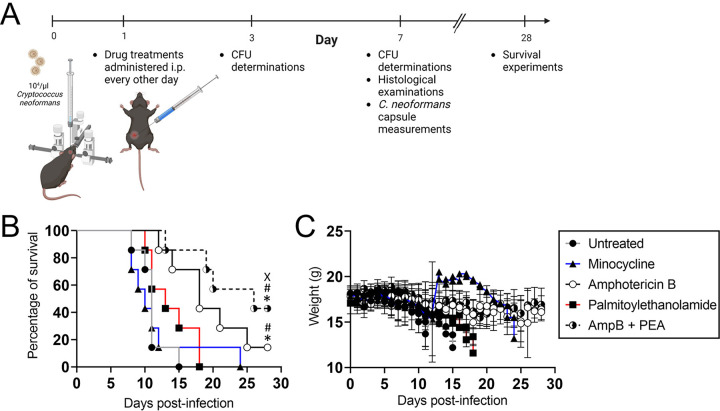
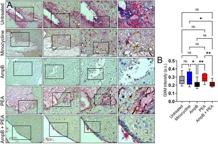
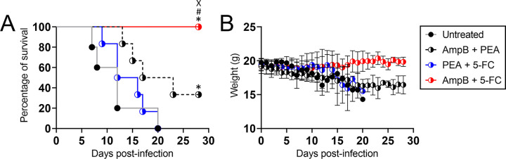
Cryptococcus neoformans ( Cn ) is an encapsulated neurotropic fungal pathogen and the causative agent of cryptococcal meningoencephalitis (CME) in humans. Recommended treatment for CME is Amphotericin B (AmpB) and 5-fluorocytosine (5-FC). Though effective, AmpB has displayed numerous adverse side effects due to its potency and nephrotoxicity, prompting investigation into alternative treatments. Palmitoylethanolamide (PEA) is an immunomodulatory compound capable of promoting neuroprotection and reducing inflammation. To investigate the efficacy of PEA as a therapeutic alternative for CME, we intracerebrally infected mice with Cn and treated them with PEA or AmpB alone or in combination. Our results demonstrate that PEA alone does not significantly prolong survival nor reduce fungal burden, but when combined with AmpB, PEA exerts an additive effect and promotes both survivability and fungal clearance. However, we compared this combination to traditional AmpB and 5-FC treatment in a survivability study and observed lower efficacy. Overall, our study revealed that PEA alone is not effective as an antifungal agent in the treatment of CME. Importantly, we describe the therapeutic capability of PEA in the context of Cn infection and show that its immunomodulatory properties may confer limited protection when combined with an effective fungicidal agent.


Conflict of interest statement

CONFLICT OF INTEREST: The authors declare no conflict of interest.

Figures

Fig. 1.
Fig. 1.. Palmitoylethanolamide (PEA) treatment prolongs survival of C57BL/6 mice infected with C. neoformans (Cn) when combined with Amphotericin B (AmpB).
(A) Experimental timeline for the intracerebral (i.c.) Cn infection and every other day intraperitoneal (i.p.) treatment model used in this study. Mice (n = 7 animals per group) were infected with 104 Cn strain H99 and administered treatments (e.g., saline (untreated), minocycline, AmpB, PEA, and AmpB + PEA) by i.p. injection every other day. Then, survival studies and colony forming units (CFU) determinations, histopathology, and microscopy were performed 3- and 7-days post-infection (dpi). The diagram was created with BioRender.com by Melissa E. Munzen. (B) Survival differences of C57BL/6 mice i.c. infected. Significance (P < 0.05) was calculated by log-rank (Mantel-Cox) analysis. *, #, and X indicate statistically different than untreated, minocycline-, and PEA-treatment groups, respectively. (C) Body weight was monitored for changes and development of clinical symptoms indicative of mice nearing endpoint. Each time point corresponds to mean weight and error bars denote standard deviations (SDs).
Fig. 2.
Fig. 2.. PEA and AmpB combination reduces cryptococcal burden in brains of mice 7 days post-infection (dpi).
(A) Periodic acid-Schiff-stained 4 μm brain sections indicating infection by Cn in C57BL/6 mice (n = 3 mice per group). Mouse brains were excised 7-dpi. Representative 10X (left panel), 20X (left center panel), 40X (right center panel), and 100X (right panel) magnifications (red-stained Cn cell wall; scale bar: 50 μm) are shown. Panel images are a magnification of the black rectangle in the corresponding left-stained section to display tissue morphology surrounding cryptococcoma in each treatment group. Arrows indicate cryptococci. (B) Fungal burden (CFU) in brains collected from Cn H99-infected mice (n = 6 mice per group) at 3- and 7-dpi. Quantification of viable yeast cells from infected animals were determined by CFU counting from two dilutions per mouse (n = 6 plates per animal) in phosphate-buffered saline (PBS). CFU determinations are based on detectable colonies at the defined concentrations in PBS (for 3-dpi, 1:1,000 and 1:10,000; for 7-dpi, 1: 10,000 and 1:50,000). Each symbol represents a single CFU determination (n = 36 plates per group). Bars and error bars denote means and SDs, respectively. Significance (****, P < 0.0001; ***, P < 0.001) was calculated by one-way analysis of variance (ANOVA) and adjusted using Tukey’s post hoc analysis. ns denotes comparisons which are not statistically significant.
Fig. 3.
Fig. 3.. Robust glial cell responses were observed around the brain region of infection of mice treated with AmpB and PEA combination.
Hematoxylin & eosin (7-dpi)-stained brain sections (4 μm thickness) from i.c. infected mice with Cn H99 (n = 3 per group) and treated with saline (untreated), minocycline, AmpB, PEA, or AmpB + PEA. Representative 10X (left panel), 20X (left center panel), 40X (right center panel), and 100X (right panel) magnifications images are shown. Arrows indicate histological changes described in the result section text. Scale bars: 50 μm.
Fig. 4.
Fig. 4.. C57BL/6 mice treated with AmpB or AmpB + PEA showed significant reduction in Cn glucuronoxylomannan (GXM) secretion.
(A) Representative images of brain tissue sections (7-dpi) from Cn H99-infected mice treated with saline (untreated), minocycline, AmpB, PEA, or AmpB + PEA co-stained with GXM-specific monoclonal antibody (mAb 18B7; red-pink) and ionized calcium binding adaptor molecule-1 (Iba-1; brown) marker for microglia. Representative 10X (left panel), 20X (left center panel), 40X (right center panel), and 100X (right panel) magnifications are shown. Panel images are a magnification of the black rectangle in the corresponding left-stained section to display tissue morphology surrounding cryptococcoma in each treatment group. Arrow indicates plugs adhered to the ependymal cells. Scale bars: 50 μm. (B) Quantification of GXM intensity. Regions of GXM release were measured (n = 15 fields per group). Boxes and whiskers denote means and SDs, respectively. Significance (**, P < 0.01; *, P < 0.05) was calculated by one-way ANOVA and adjusted using Tukey’s post hoc analysis. ns denotes comparisons which are not statistically significant.
Fig. 5.
Fig. 5.. Brains from C57BL/6 mice treated with AmpB or AmpB + PEA exhibit cryptococci with considerable capsule size decrease.
(A) Images of brain homogenates (7-dpi) from mice i.c. infected with 104 Cn cells and treated with saline (untreated), minocycline, AmpB, PEA, or AmpB + PEA. Fungal cells were stained with India Ink. Each image was examined by light microscopy using a Leica DMi8 inverted microscope and images captured with a Leica DFC7000 digital camera using LAS X digital imaging software. (B) Capsule volume (V = 4/3 π(R2-r2) for Cn cells in brain homogenates from each group was calculated using Leica LAS X software. Brain homogenates from 3 mice per group were analyzed, and ≥ 200 cells were measured. Bars and error bars denote means and SDs, respectively. Significance (*, P < 0.05) was calculated by one-way ANOVA and adjusted using Tukey’s post hoc analysis. ns denotes comparisons which are not statistically significant.
Fig. 6.
Fig. 6.. Differential microglial morphology in brain tissue infected with Cn after treatment with AmpB, PEA, or combination.
(A) Representative images of brain tissue sections (7-dpi) from Cn H99-infected mice (n = 3 mice per group) treated with saline (untreated), minocycline, AmpB, PEA, or AmpB + PEA co-stained with mAb 18B7 (GXM, red-pink) and Iba-1 (microglia, brown). Representative 10X (left panel), 20X (left center panel), 40X (right center panel), and 100X (right panel) magnifications are shown. Panel images are a magnification of the black rectangle in the corresponding left-stained section to display microglia morphological changes near a cryptococcoma in each treatment group. Scale bars: 50 μm. (B) Pie charts showing the percentage of microglial morphology distribution (e.g., activated, ramified, dystrophic, phagocytic/amoeboid, and rod-shaped cells) in brain tissue during infection with Cn H99 and different treatments. Microglial phenotype abundance was visualized using light microscopy and classified according to their morphology.
Fig. 7.
Fig. 7.. Combination of AmpB and PEA prolongs survival of C57BL/6 mice, although not with efficacy comparable to AmpB and 5-Fluorocytosine (5-FC).
(A) Survival differences of C57BL/6 mice i.c. infected with 104 Cn strain H99 (n = 6 per group) and every other day given i.p. treatment with saline (untreated), AmpB + PEA, PEA + 5-FC, or AmpB + 5-FC. Significance (P < 0.05) was calculated by log-rank (Mantel-Cox) analysis. *, #, and X indicate statistically different than untreated, AmpB + PEA−, and PEA + 5-FC-treatment groups, respectively. (B) Body weight was monitored for changes and development of clinical symptoms indicative of mice nearing endpoint. Each time point corresponds to mean weight and error bars denote SDs.

Similar articles

References

    1. Kabir Z, Cunningham C. 2022. The global burden of cryptococcosis-a neglected tropical disease? Lancet Infect Dis 22:1658–1660. - PubMed
    1. Rajasingham R, Smith RM, Park BJ, Jarvis JN, Govender NP, Chiller TM, Denning DW, Loyse A, Boulware DR. 2017. Global burden of disease of HIV-associated cryptococcal meningitis: an updated analysis. Lancet Infect Dis 17:873–881. - PMC - PubMed
    1. Rajasingham R, Govender NP, Jordan A, Loyse A, Shroufi A, Denning DW, Meya DB, Chiller TM, Boulware DR. 2022. The global burden of HIV-associated cryptococcal infection in adults in 2020: a modelling analysis. Lancet Infect Dis 22:1748–1755. - PMC - PubMed
    1. Williamson PR, Jarvis JN, Panackal AA, Fisher MC, Molloy SF, Loyse A, Harrison TS. 2017. Cryptococcal meningitis: epidemiology, immunology, diagnosis and therapy. Nat Rev Neurol 13:13–24. - PubMed
    1. Kneale M, Bartholomew JS, Davies E, Denning DW. 2016. Global access to antifungal therapy and its variable cost. J Antimicrob Chemother 71:3599–3606. - PubMed

Publication types