

Respiratory muscle ultrasonography evaluation and its clinical application in stroke patients: A review
- PMID: 37090789
- PMCID: PMC10115993
- DOI: 10.3389/fnins.2023.1132335
Respiratory muscle ultrasonography evaluation and its clinical application in stroke patients: A review
Abstract
Background: Respiratory muscle ultrasound is a widely available, highly feasible technique that can be used to study the contribution of the individual respiratory muscles related to respiratory dysfunction. Stroke disrupts multiple functions, and the respiratory function is often significantly decreased in stroke patients.
Method: A search of the MEDLINE, Web of Science, and PubMed databases was conducted. We identified studies measuring respiratory muscles in healthy and patients by ultrasonography. Two reviewers independently extracted and documented data regarding to the criteria. Data were extracted including participant demographics, ultrasonography evaluation protocol, subject population, reference values, etc.
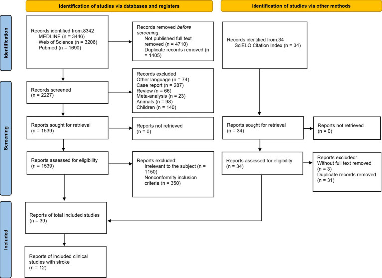
Result: A total of 1954 participants from 39 studies were included. Among them, there were 1,135 participants from 19 studies on diaphragm, 259 participants from 6 studies on extra-diaphragmatic inspiratory muscles, and 560 participants from 14 studies on abdominal expiratory muscles. The ultrasonic evaluation of diaphragm and abdominal expiratory muscle thickness had a relatively typically approach, while, extra-diaphragmatic inspiratory muscles were mainly used in ICU that lack of a consistent paradigm.
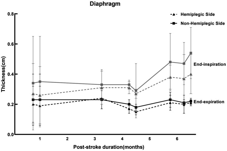
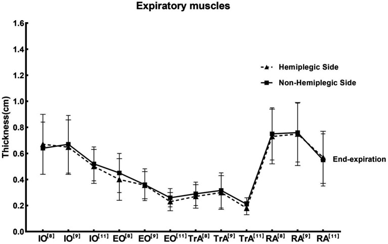
Conclusion: Diaphragm and expiratory muscle ultrasound has been widely used in the assessment of respiratory muscle function. On the contrary, there is not enough evidence to assess extra-diaphragmatic inspiratory muscles by ultrasound. In addition, the thickness of the diaphragm on the hemiplegic side was lower than that on the non-hemiplegic side in stroke patients. For internal oblique muscle (IO), rectus abdominis muscle (RA), transversus abdominis muscle (TrA), and external oblique muscle (EO), most studies showed that the thickness on the hemiplegic side was lower than that on the non-hemiplegic side.Clinical Trial Registration: The protocol of this review was registered in the PROSPERO database (CRD42022352901).
Keywords: diaphragm; evaluation; respiratory muscle; stroke; ultrasonography.
Copyright © 2023 Liu, Yang and Jia.
Conflict of interest statement
The authors declare that the research was conducted in the absence of any commercial or financial relationships that could be construed as a potential conflict of interest.
Figures
References
-
- Abuín-Porras V., Maldonado-Tello P., de la Cueva-Reguera M., Rodríguez-Sanz D., Calvo-Lobo C., López-López D., et al. (2020). Comparison of lateral abdominal musculature activation during expiration with an expiratory flow control device versus the abdominal drawing-in maneuver in healthy women: a cross-sectional observational pilot study. Medicina 56:84. doi: 10.3390/medicina56020084, PMID: - DOI - PMC - PubMed
Publication types
LinkOut - more resources
Full Text Sources