

Case Reports
doi: 10.1055/s-0041-1723959.
eCollection 2023 Jun.
Eye of the Tiger: Looking Beyond Neurodegeneration with Brain Iron Accumulation Disorders
Affiliations
- PMID: 37090832
- PMCID: PMC10118706
- DOI: 10.1055/s-0041-1723959
Item in Clipboard
Case Reports
Eye of the Tiger: Looking Beyond Neurodegeneration with Brain Iron Accumulation Disorders
J Pediatr Genet.
.
Abstract
The "eye-of-the-tiger" sign in brain magnetic resonance imaging (MRI) is typically associated with neurodegeneration with brain iron accumulation disorders, especially pantothenate kinase-associated neurodegeneration. However, very similar neuroimaging findings may be seen in other neurodegenerative disorders involving the basal ganglia. We report here a patient with fucosidosis who had MRI brain findings closely resembling the "eye-of-the-tiger" sign.
Keywords: eye-of-the-tiger sign; fucosidosis; neurodegeneration with brain iron accumulation.
Thieme. All rights reserved.
Conflict of interest statement
Conflict of Interest None declared.
Figures
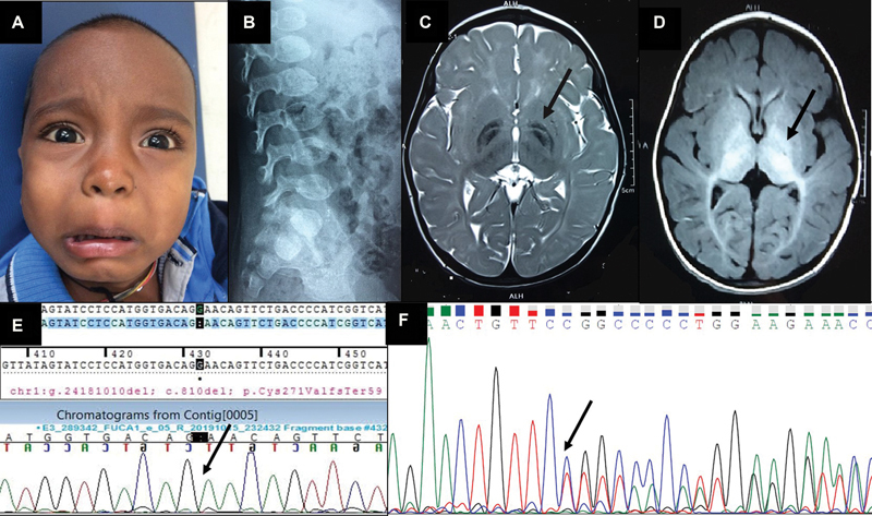
(
A
) Close-up of the child's face showing the coarse facial features, mild ocular hypertelorism, and broad nasal bridge and thick lips. (
B
) Lateral radiograph of the lumbosacral spine showing the ovoid vertebrae and the anterior-inferior beaking indicative of dysostosis multiplex. (
C
) T2-weighted axial image of MRI brain showing bilaterally symmetrical hypointensity of both the globi pallidi with a central streak of hyperintensity between the medial and lateral segments of the globi pallidi, resembling the “eye-of-the-tiger” sign (pointed out by the arrow), along with symmetric hyperintensities in bilateral cerebral white matter. (
D
) T1-weighted axial image of MRI brain showing bilaterally symmetrical hyperintensity of globi pallidi, substantia nigra, and subthalamic nuclei (pointed out by the arrow). (
E
) Sanger sequence chromatogram of the proband showing the homozygous c.810del variant in the
FUCA1
gene (the position of the single nucleotide deletion is marked with the arrow). (
F
) Sanger sequence chromatogram of the mother showing the heterozygous c.810del variant in the
FUCA1
gene (the position of the single nucleotide deletion is marked with the arrow).
Similar articles
-
Type 1 neurodegeneration with brain iron accumulation: a case report.J Med Case Rep. 2022 Jun 3;16(1):217. doi: 10.1186/s13256-022-03430-7. J Med Case Rep. 2022. PMID: 35655240 Free PMC article.
-
Pantothenate kinase 2 mutation with eye-of-the-tiger sign on magnetic resonance imaging in three siblings.Iran J Neurol. 2012;11(4):155-8. Iran J Neurol. 2012. PMID: 24250886 Free PMC article.
-
Eye-of-the-tiger Sign in Neurodegeneration with Brain Iron Accumulation.Cureus. 2019 Jun 18;11(6):e4936. doi: 10.7759/cureus.4936. Cureus. 2019. PMID: 31431841 Free PMC article.
-
Update on neurodegeneration with brain iron accumulation.Neurol Neurochir Pol. 2014;48(3):206-13. doi: 10.1016/j.pjnns.2014.05.001. Epub 2014 May 17. Neurol Neurochir Pol. 2014. PMID: 24981186 Review.
-
NBIA Syndromes: A Step Forward from the Previous Knowledge.Neurol India. 2021 Sep-Oct;69(5):1380-1388. doi: 10.4103/0028-3886.329603. Neurol India. 2021. PMID: 34747818 Review.
Cited by
-
Early Severe Cortical Involvement and Novel FUCA1 Mutations in a Pediatric Fucosidosis Case.Mol Genet Genomic Med. 2025 Feb;13(2):e70070. doi: 10.1002/mgg3.70070. Mol Genet Genomic Med. 2025. PMID: 39865383 Free PMC article.
References
-
- ACMG Laboratory Quality Assurance Committee . Richards S, Aziz N, Bale S. Standards and guidelines for the interpretation of sequence variants: a joint consensus recommendation of the American College of Medical Genetics and Genomics and the Association for Molecular Pathology. Genet Med. 2015;17(05):405–424. - PMC - PubMed
-
- George H T. 8th ed. New York: McGraw-Hill; 2001. Disorders of glycoprotein degradation: α-mannosidosis, β-mannosidosis, α-fucosidosis and sialidosis; pp. 3507–3533.
-
- Sheth J, Mistri M, Bhavsar R. Lysosomal storage disorders in Indian children with Neuroregression attending a genetic center. Indian Pediatr. 2015;52(12):1029–1033. - PubMed
-
- Wali G, Wali G M, Sue C M, Kumar K R. A novel homozygous mutation in the FUCA1 gene highlighting fucosidosis as a cause of dystonia: case report and literature review. Neuropediatrics. 2019;50(04):248–252. - PubMed
Publication types
LinkOut - more resources
Full Text Sources
