. 2023 Mar 23;26(4):106470.
doi: 10.1016/j.isci.2023.106470. eCollection 2023 Apr 21.

An inducible explant model of osteoclast-osteoprogenitor coordination in exacerbated osteoclastogenesis

Affiliations

An inducible explant model of osteoclast-osteoprogenitor coordination in exacerbated osteoclastogenesis

Jarred M Whitlock et al. iScience. .

Abstract

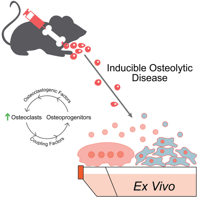
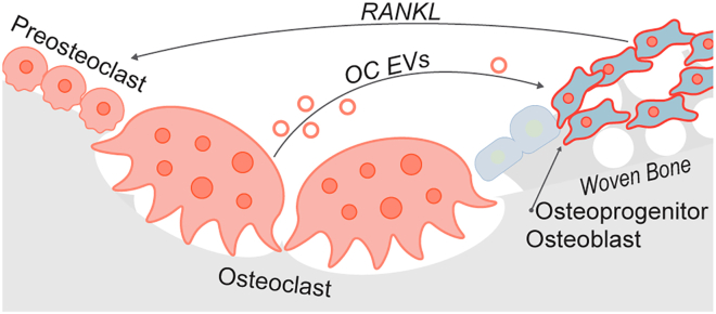
Elucidating a basic blueprint of osteoclast-osteoblast coordination in skeletal remodeling and understanding how this coordination breaks down with age and disease is essential for addressing the growing skeletal health problem in our aging population. The paucity of simple, activatable, biologically relevant models of osteoclast-osteoblast coordination has hindered our understanding of how skeletal remolding is regulated. Here, we describe an inducible ex vivo model of osteoclast-osteoblast progenitor coordination. Induction activates the release of osteoclastogenic factors from osteoprogenitors, which elicits the differentiation and fusion of neighboring preosteoclasts. In turn, multinucleated osteoclasts release soluble coupling factors, RANK+ extracellular vesicles and promote osteoprogenitor proliferation, recapitulating aspects of perturbed coordination in diseases underpinned by excessive osteoclast formation. We expect this model to expedite the investigation of cell-cell fusion, osteoclast-osteoblast progenitor coordination, and extracellular vesicle signaling during bone remodeling and offer a powerful tool for evaluating signaling cascades and novel therapeutic interventions in osteoclast-linked skeletal disease.

Keywords: Biological sciences; Cell biology; Stem cells research.


Conflict of interest statement

The authors declare no competing interests.

Figures

None
Graphical abstract
Figure 1
Figure 1
Induction of sR201C in osteoprogenitors induces osteoclast formation in marrow cultures (A) A cartoon schematic the genetic tools utilized and our approach for implementing them to model oteoprogenitor-osteoclast signaling/coordination in the formation of osteoclasts. (B) A representative Western blot evaluating Gαs expression with (Dox.) or without (Unind.) 4 days induction. (C) ELISA evaluation of soluble RANKL in the culture media of marrow explants with (Dox.) or without (Unind.) 4 days induction. (n = 7). (D) ELISA evaluation of soluble IL-6 in the culture media of marrow explants with (Dox.) or without (Unind.) 4 days induction. (n = 4). (E) ELISA evaluation of soluble M-CSF in the culture media of marrow explants with (Dox.) or without (Unind.) 4 days induction. (n = 5). (F) Representative color brightfield images of marrow explants from GαsR201C following 4 days with (Dox.), without (Unind.) or with induction in the presence of OPG (Dox.+OPG). Arrow denotes x-gal staining of the nucleus of an induced osteoprogenitor, Arrowhead denotes a multinucleated, TRAP+ osteoclast. Error bars = ±SEM. Significance was assessed via paired t-test, where ∗p=<0.05, ∗∗p=<0.01, ∗∗∗p=<0.001.
Figure 2
Figure 2
Dox. induction elicits osteoclast formation (A) Representative images of ex vivo marrow cultures following Dox. induction (Gray = Phalloidin-Alexa488, Cyan = Hoechst). (B) Quantification of the number of fusion events over time following addition of Dox. Each time course was normalized to the number of fusion events observed on day 5. (n = 3). (C) qPCR evaluation of the osteoclastogenesis transcription factors Nfatc1 and cFos under uninduced (black), Dox. induced (gray) or Dox. induced +100 ng/mL OPG (white) conditions. Fold expression is relative to Actb RNA. (n = 3). (D) Quantification of the number of fusion events in ex vivo marrow cultures treated as in (C). (n = 3). (E) Representative Western blot evaluation of the steady-state level of the osteoclast resorption enzyme CTSK in ex vivo marrow cultures treated as in (C). (F) Representative immunofluorescence images of the pan macrophage lineage marker CD68 (green), the osteoclast sorting nexin SNX10 and Hoechst in ex vivo marrow cultures treated as in (C). (G) Quantification of bone resorption by ex vivo marrow cultures treated as in (C). In (A and D), Arrowheads = multinucleated osteoclasts, Arrows = proliferative preosteoblasts. Error bars = ±SEM. Statistical significance was assessed via paired t-test, where ∗p=<0.05, ∗∗p=<0.01, ∗∗∗p=<0.001.
Figure 3
Figure 3
Osteoclast formation in ex vivo cultures elicits osteoprogenitor proliferation (A) Representative phase contrast images of TRAP stained marrow explants following 4 days with (Dox.), without (Unind.) or with induction in the presence of OPG (Dox.+OPG). Arrow denotes lesion of cells, Arrowheads denote TRAP+, multinucleated osteoclasts, which appear dark in phase contrast. (B) ELISA evaluation of soluble LIF in the culture media of marrow explants without (Unind.), with 4 days induction (Dox.) or 4 days induction and 100 ng/mL OPG (OPG). (n = 6 for OPG or n = 7 for Unind. and Dox.). (C) Representative immunofluorescence images of the preosteoblast marker Runx2, the proliferation marker Ki67 and the nuclear stain Hoechst in cultures treated as in (A). (D) Quantification of the fraction of Runx2+ preosteoblasts that exhibit staining for the proliferation marker Ki67. (n = 3). (E) qPCR evaluation of the osteogenic markers Sp7, Dlx5 and Bglap under uninduced (black), Dox. induced (gray) or Dox. induced +100 ng/mL OPG (white) conditions. Fold expression is relative to 18s rRNA. (n = 5). (F) Representative immunofluorescence images of the preosteoblast marker Runx2, the proliferation marker Ki67 and the nuclear stain Hoechst in MC3T3-E1 clone 14 preosteoblasts treated with (OPG) or without (control) 100 ng/mL OPG. (G) Quantification of the fraction of Runx2+ preosteoblasts that exhibit staining for the proliferation marker Ki67 treated as in (F). (n = 3 for clone 4(circle), n = 3 for clone 14(square). Bar denotes summary average of both clones 4 and 14). (H) qPCR evaluation of the osteogenic markers Sp7, Dlx5 and Bglap in clones 4 and 14 under control (black), 100 ng/mL OPG (white) conditions. Fold expression is relative to 18s rRNA. (n = 3) Error bars = ±SEM. Significance was assessed via paired t-test, where ∗p=<0.05, ∗∗p=<0.01, ∗∗∗p=<0.001.
Figure 4
Figure 4
Osteoclast formation in ex vivo marrow cultures is accompanied by EV release (A) An illustrated schematic depicting an approach for enriching an EV fraction from uninduced, Doxy. induced and Doxy. induced +100 ng/mL OPG ex vivo marrow cultures (left). A representative Western blot evaluating EV enriched fractions for the osteoclast membrane receptor RANK and the exosome marker TSG101 (right). (B) Quantification of RANK signal in representative Western blots of EV enriched fractions, as in (A). (n = 3). (C) Quantification of TSG101 signal in representative Western blots of EV enriched fractions, as in (A). (n = 5) Error bars = ±SEM. Significance was assessed via paired t-test, where ∗p=<0.05, ∗∗p=<0.01, ∗∗∗p=<0.001.
Figure 5
Figure 5
Ex vivo FD marrow explants as a model of osteoclast formation and osteoclast-osteoprogenitor coordination An illustration of the biological process modeled in our ex vivo marrow cultures. Doxycycline induction elicits osteoprogenitor expression of GαsR201C and release of RANKL. RANKL binding initiates osteoclastogenesis and the formation of multinucleated, bone resorbing osteoclasts. Osteoclasts release EVs that correlate with preosteoblast proliferation. Peach cells are of the monocyte>osteoclast lineage. Blue cells are of the skeletal stem cell>osteoblast lineage.

Similar articles

Cited by

References

    1. Feng X., McDonald J.M. Disorders of bone remodeling. Annu. Rev. Pathol. 2011;6:121–145. doi: 10.1146/annurev-pathol-011110-130203. - DOI - PMC - PubMed
    1. Raggatt L.J., Partridge N.C. Cellular and molecular mechanisms of bone remodeling. J. Biol. Chem. 2010;285:25103–25108. doi: 10.1074/jbc.R109.041087. - DOI - PMC - PubMed
    1. Kodama H., Nose M., Niida S., Yamasaki A. Essential role of macrophage colony-stimulating factor in the osteoclast differentiation supported by stromal cells. J. Exp. Med. 1991;173:1291–1294. doi: 10.1084/jem.173.5.1291. - DOI - PMC - PubMed
    1. Nakashima T., Hayashi M., Fukunaga T., Kurata K., Oh-Hora M., Feng J.Q., Bonewald L.F., Kodama T., Wutz A., Wagner E.F., et al. Evidence for osteocyte regulation of bone homeostasis through RANKL expression. Nat. Med. 2011;17:1231–1234. doi: 10.1038/nm.2452. - DOI - PubMed
    1. Li D., Liu J., Guo B., Liang C., Dang L., Lu C., He X., Cheung H.Y.S., Xu L., Lu C., et al. Osteoclast-derived exosomal miR-214-3p inhibits osteoblastic bone formation. Nat. Commun. 2016;7:10872. doi: 10.1038/ncomms10872. - DOI - PMC - PubMed

LinkOut - more resources