. 2023 Apr 6:11:1165465.
doi: 10.3389/fbioe.2023.1165465. eCollection 2023.

Recombinant production, purification, and biochemical characterization of a novel L-lactate dehydrogenase from Bacillus cereus NRC1 and inhibition study of mangiferin

Affiliations

Recombinant production, purification, and biochemical characterization of a novel L-lactate dehydrogenase from Bacillus cereus NRC1 and inhibition study of mangiferin

Sayed S Esa et al. Front Bioeng Biotechnol. .

Abstract

Lactate dehydrogenase (LDH, EC 1.1.1.27) is one of the vital glycolytic conditions, especially during anaerobic conditions. It is a significant diagnostic, prognostic, and monitoring biomarker parameter. A 950-bp DNA fragment containing the gene (LDH) encoding LDH was amplified from Bacillus cereus NRC1. The deduced amino acid sequence reveals that B. cereus LDH (Bc-LDH) is highly homologous to the LDHs of Bacillus organisms. All LDH enzymes have a significant degree of conservation in their active site and several additional domains with unidentified functions. The gene for LDH, which catalyzes lactate synthesis, was cloned, sequenced (accession number: LC706200.1), and expressed in Escherichia coli BL21 (DE3). In this investigation, Bc-LDH was purified to homogeneity with a specific activity of 22.7 units/mg protein and a molecular weight of 35 kDa. It works optimally at pH 8.0. The purified enzyme was inhibited by FeCl2, CuCl2, ZnCl2, and NiCl, whereas CoCl2 was found to boost the activity of Bc-LDH. The molecular docking of the 3D model of the Bc-LDH structure with a natural inhibitor, mangiferin, demonstrated excellent LDH inhibition, with a free binding energy of -10.2 kcal/mol. Moreover, mangiferin is a potent Bc-LDH inhibitor that inhibits Bc-LDH competitively and has one binding site with a Ki value of 0.075 mM. The LDH-mangiferin interaction exhibits a low RMSF value (>1.5 Å), indicating a stable contact at the residues. This study will pave the way for more studies to improve the understanding of mangiferin, which could be considered an intriguing candidate for creating novel and improved LDH inhibitors.

Keywords: Bacillus cereus; characterization; cloning; lactate dehydrogenase; mangiferin; molecular docking.


Conflict of interest statement

The authors declare that the research was conducted in the absence of any commercial or financial relationships that could be construed as a potential conflict of interest.

Figures

FIGURE 1
FIGURE 1
(A) Bacterial identification PCR—Identified by 16S rRNA Sequences. (B) Phylogenetic analysis produced as a result of the repeated alignment of the 16s rRNA gene sequence of Bacillus cereus NRC1 with the sequences of other Bacillus strains located in the GenBank database.
FIGURE 2
FIGURE 2
(A) PCR amplification of 0.95 kb Bc-LDH gene from the chromosomal DNA of Bacillus cereus NRC1. (B) Complete nucleotide sequence of the Bc-LDH gene from B. cereus open reading frame (ORF) nucleotide sequence. (C) Complete amino acid sequences and composition of the Bc-LDH gene from B. cereus NRC1. (D) SDS-PAGE showing a single band with a molecular weight of 35 kDa.
FIGURE 3
FIGURE 3
(A) LDH gene sequences from Bacillus cereus NRC1 and other organisms aligned based on their recognized amino acid sequences. The following is a list of accession numbers for the other sequences: LDH-NRC1, B. cereus ATCC, Bacillus thuringiensis, Bacillus anthracis, and Bacillus manliponensis LDH, whose GenBank accession numbers are BDI00612.1, Q81EP4, Q6HK31, Q81RW4, and A0A073K0E2, respectively. Identity is indicated by a “*” symbol and similarity is indicated by a “:” symbol. Also, the red box shows active site positions and the black box represents the signal peptide region (1–17 residues). (B) Percent identity matrix of LDH protein of B. cereus NRC1 and other organisms.
FIGURE 4
FIGURE 4
Phylogenetic tree of Bc-LDH proteins from Bacillus cereus strain NRC1 and other L-lactate dehydrogenase proteins inferred using the neighbor-joining method (MEGA X).
FIGURE 5
FIGURE 5
Elution profile of Bc-LDH: protein UV absorption and LDH activity. (A) Elution profile on DEAE-cellulose, pH 7.6. (B) Elution profile of Bc-LDH DEAE-cellulose fractions on CM-cellulose column, pH 5.5. (C) Bc-LDH CM-cellulose fractions on Sephacryl S-300 column, pH 7.6. (D) 7% native PAGE of LDH pattern. (E) LDH isoenzyme activity.
FIGURE 6
FIGURE 6
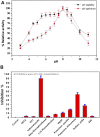
(A) Impact of pH on the purified LDH, using 0.05 M of pHs ranging from 3.0 to 11. (B) Effect of inhibitors on the purified LDH from Bacillus cereus.
FIGURE 7
FIGURE 7
Structural alignment of the LDH gene from Bacillus cereus with B. cereus (PDB 4LMR_A). The actual secondary structure of the templates and the predicted secondary structure of the lactate dehydrogenase gene are shown above the alignment. The symbols η, α, and ß, respectively, stand for the secondary structure components 310 helices, a-helices, and ß-strands.
FIGURE 8
FIGURE 8
(A, B) Superimposition of model of LDH protein from Bacillus cereus (predicted model) and template B. cereus (PDB 4LMR_A) through cartoon representation. (C, D) Ramachandran plot for template B. cereus (PDB 4LMR_A) and model of Bc-LDH protein.
FIGURE 9
FIGURE 9
(A) 2D interaction: revealing hydrogen bond between ligands interacted with an amino acid (green dash line). (B) Complex interaction: showing binding of mangiferin with Bc-LDH displaying the most effective binding mode in the protein cavity (active site displayed by yellow color). (C) 3D interaction: showing the interacted amino acid residues (with ligand as color sticks).
FIGURE 10
FIGURE 10
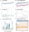
(A) RMSD plot of Bc-LDH protein, (B) RMSD plot of Bc-LDH-mangiferin complex, (C) RMSF plot of Bc-LDH protein, (D) RMSF plot of mangiferin, (E, F) Bc-LDH-mangiferin complex contact interactions.
FIGURE 11
FIGURE 11
(A) RMSD plot of mangiferin, (B) rGyr of mangiferin, (C) intraHB within mangiferin molecule, (D) MolSA of mangiferin, (E) SASA of mangiferin, and (F) PSA of mangiferin.
FIGURE 12
FIGURE 12
(A) Lineweaver-Burk plots reveal the LDH inhibition type using mangiferin. (B) Estimation of the inhibition constant (Ki) for LDH inhibition using mangiferin.

References

    1. Abosereh N. A., Salim R. G., El-Sayed A. F., Hammad M. A., Elsayed G. M. (2022). In-vitro biodegradation of Glyphosate using genetically improved bacterial isolates from highly polluted wastewater. Egypt. J. Chem. 65, 0–681. 10.21608/EJCHEM.2022.141571.6194 - DOI
    1. Andreeßen C., Wolf N., Cramer B., Humpf H.-U., Steinbüchel A. (2018). In vitro biosynthesis of 3-mercaptolactate by lactate dehydrogenases. Enzyme Microb. Technol. 108, 1–10. 10.1016/j.enzmictec.2017.08.005 - DOI - PubMed
    1. Andrews P. (1964). Estimation of the molecular weights of proteins by Sephadex gel-filtration. Biochem. J. 91, 222–233. 10.1042/bj0910222 - DOI - PMC - PubMed
    1. Aziz N., Kim M. Y., Cho J. Y. (2018). Anti-inflammatory effects of luteolin: A review of in vitro, in vivo, and in silico studies. J. Ethnopharmacol. 225, 342–358. 10.1016/j.jep.2018.05.019 - DOI - PubMed
    1. Banu M. J., Nellaiappan K., Dhandayuthapani S. (1992). Mitochondrial malate dehydrogenase and malic enzyme of a filarial worm setaria digitata: Some properties and effects of drugs and herbal extracts. Jpn. J. Med. Sci. Biol. 45, 137–150. 10.7883/yoken1952.45.137 - DOI - PubMed

LinkOut - more resources