

Corneal optical density: Structural basis, measurements, influencing factors, and roles in refractive surgery
- PMID: 37091331
- PMCID: PMC10117965
- DOI: 10.3389/fbioe.2023.1144455
Corneal optical density: Structural basis, measurements, influencing factors, and roles in refractive surgery
Abstract
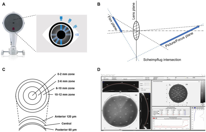
The cornea is the main refractive medium of the human eye, and its clarity is critical to visual acuity. Corneal optical density (COD) is an important index to describe corneal transparency. Intact corneal epithelial and endothelial cells, regular arrangement of collagen fibers in the stroma, and normal substance metabolism are all integral for the cornea to maintain its transparency. In the last two decades, the Pentacam Scheimpflug imaging system has emerged as a breakthrough for the measurement of COD (also called corneal densitometry). It has been found that a wide variety of factors such as age, refractive status, and corneal diseases can affect COD. Different corneal refractive surgery methods also change COD in different corneal regions and layers and affect visual acuity following the surgery. Thus, COD has gradually become a significant indicator to evaluate corneal health, one on which the attention of clinicians has been increasingly focused.
Keywords: FS-LASIK; SMILE; corneal densitometry; corneal optical density; pentacam scheimpflug imaging system.
Copyright © 2023 He, Ma, Zeng and Ma.
Conflict of interest statement
The authors declare that the research was conducted in the absence of any commercial or financial relationships that could be construed as a potential conflict of interest. The reviewer TH declared a shared parent affiliation with the authors BM and JZ to the handling editor at the time of review.
Figures
Similar articles
-
[Clinical observation on corneal transparency after small incision lenticule extraction surgery].Zhonghua Yan Ke Za Zhi. 2018 Jan 11;54(1):27-32. doi: 10.3760/cma.j.issn.0412-4081.2018.01.006. Zhonghua Yan Ke Za Zhi. 2018. PMID: 29429284 Chinese.
-
In vivo confocal microscopy of sub-basal corneal nerves and corneal densitometry after three kinds of refractive procedures for high myopia.Int Ophthalmol. 2023 Mar;43(3):925-935. doi: 10.1007/s10792-022-02494-0. Epub 2022 Sep 25. Int Ophthalmol. 2023. PMID: 36153757
-
Corneal Densitometry after Femtosecond Laser-Assisted In Situ Keratomileusis (Fs-LASIK) and Small Incision Lenticule Extraction (SMILE).Curr Eye Res. 2018 May;43(5):605-610. doi: 10.1080/02713683.2018.1431288. Epub 2018 Mar 14. Curr Eye Res. 2018. PMID: 29537886
-
[Corneal densitometry : Value for keratoconus diagnostics].Ophthalmologe. 2018 Sep;115(9):737-743. doi: 10.1007/s00347-018-0667-3. Ophthalmologe. 2018. PMID: 29468296 Review. German.
-
[The cornea: stasis and dynamics].Nippon Ganka Gakkai Zasshi. 2008 Mar;112(3):179-212; discussion 213. Nippon Ganka Gakkai Zasshi. 2008. PMID: 18411711 Review. Japanese.
Cited by
-
Impacts and Correlations on Corneal Biomechanics, Corneal Optical Density and Intraocular Pressure after Cataract Surgery.Diagnostics (Basel). 2024 Jul 18;14(14):1557. doi: 10.3390/diagnostics14141557. Diagnostics (Basel). 2024. PMID: 39061693 Free PMC article.
-
A 6-Month Follow-Up Comparative Study of Single-Step Transepithelial Photorefractive Keratectomy (Trans-PRK) Using the StreamLight Software with and without Epithelial Thickness Customization.Clin Ophthalmol. 2024 Oct 9;18:2831-2841. doi: 10.2147/OPTH.S487627. eCollection 2024. Clin Ophthalmol. 2024. PMID: 39398468 Free PMC article.
-
Corneal Stroma Analysis and Related Ocular Manifestations in Recovered COVID-19 Patients.Invest Ophthalmol Vis Sci. 2024 May 1;65(5):14. doi: 10.1167/iovs.65.5.14. Invest Ophthalmol Vis Sci. 2024. PMID: 38713483 Free PMC article.
-
The Impact of Standard Cross-Linking on the Corneal Optical Density-Age Relationship in Keratoconus After Mechanical Stripping of the Epithelium.J Ophthalmol. 2024 Oct 15;2024:8827837. doi: 10.1155/2024/8827837. eCollection 2024. J Ophthalmol. 2024. PMID: 39444422 Free PMC article.
References
-
- Alio Del Barrio J. L., Parafita-Fernandez A., Canto-Cerdan M., Alio J. L., Teus M. (2021). Evolution of corneal thickness and optical density after laser in situ keratomileusis versus small incision lenticule extraction for myopia correction. Br. J. Ophthalmol. 105, 1656–1660. 10.1136/bjophthalmol-2020-316601 - DOI - PubMed
Publication types
LinkOut - more resources
Full Text Sources