A CS-based composite scaffold with excellent photothermal effect and its application in full-thickness skin wound healing
- PMID: 37091498
- PMCID: PMC10118997
- DOI: 10.1093/rb/rbad028
A CS-based composite scaffold with excellent photothermal effect and its application in full-thickness skin wound healing
Abstract
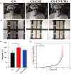
The development of natural polymer-based scaffolds with excellent biocompatibility, antibacterial activity, and blood compatibility, able to facilitate full-thickness skin wound healing, remains challenging. In this study, we have developed three chitosan (CS)-based porous scaffolds, including CS, CS/CNT (carbon nanotubes) and CS/CNT/HA (nano-hydroxyapatite, n-HA) using a freeze-drying method. All three scaffolds have a high swelling ratio, excellent antibacterial activity, outstanding cytocompatibility and blood compatibility in vitro. The introduction of CNTs exhibited an obvious increase in mechanical properties and exerts excellent photothermal response, which displays excellent healing performance as a wound dressing in mouse full-thickness skin wound model when compared to CS scaffolds. CS/CNT/HA composite scaffolds present the strongest ability to promote full-thickness cutaneous wound closure and skin regeneration, which might be ascribed to the synergistic effect of photothermal response from CNT and excellent bioactivity from n-HA. Overall, the present study indicated that CNT and n-HA can be engineered as effective constituents in wound dressings to facilitate full-thickness skin regeneration.
Keywords: carbon nanotubes; chitosan; hydroxyapatite nanoparticles; photothermal effect; skin regeneration.
© The Author(s) 2023. Published by Oxford University Press.
Figures


Similar articles
-
Mussel-inspired, antibacterial, conductive, antioxidant, injectable composite hydrogel wound dressing to promote the regeneration of infected skin.J Colloid Interface Sci. 2019 Nov 15;556:514-528. doi: 10.1016/j.jcis.2019.08.083. Epub 2019 Aug 24. J Colloid Interface Sci. 2019. PMID: 31473541
-
The improvement of periodontal tissue regeneration using a 3D-printed carbon nanotube/chitosan/sodium alginate composite scaffold.J Biomed Mater Res B Appl Biomater. 2023 Jan;111(1):73-84. doi: 10.1002/jbm.b.35133. Epub 2022 Jul 16. J Biomed Mater Res B Appl Biomater. 2023. PMID: 35841326
-
[Influence of collagen/fibroin scaffolds containing silver nanoparticles on dermal regeneration of full-thickness skin defect wound in rat].Zhonghua Shao Shang Za Zhi. 2017 Feb 20;33(2):103-110. doi: 10.3760/cma.j.issn.1009-2587.2017.02.011. Zhonghua Shao Shang Za Zhi. 2017. PMID: 28219143 Chinese.
-
Local anti-inflammatory effect and immunomodulatory activity of chitosan-based dressing in skin wound healing: A systematic review.J Clin Transl Res. 2022 Nov 9;8(6):488-498. eCollection 2022 Dec 29. J Clin Transl Res. 2022. PMID: 36451998 Free PMC article. Review.
-
Applications of Carbon Nanotubes in Bone Regenerative Medicine.Nanomaterials (Basel). 2020 Apr 2;10(4):659. doi: 10.3390/nano10040659. Nanomaterials (Basel). 2020. PMID: 32252244 Free PMC article. Review.
Cited by
-
The utilization of chitosan/Bletilla striata hydrogels to elevate anti-adhesion, anti-inflammatory and pro-angiogenesis properties of polypropylene mesh in abdominal wall repair.Regen Biomater. 2024 Apr 27;11:rbae044. doi: 10.1093/rb/rbae044. eCollection 2024. Regen Biomater. 2024. PMID: 38962115 Free PMC article.
-
In vitro and in vivo assessment of a non-animal sourced chitosan scaffold loaded with xeno-free umbilical cord mesenchymal stromal cells cultured under macromolecular crowding conditions.Biomater Biosyst. 2024 Oct 10;16:100102. doi: 10.1016/j.bbiosy.2024.100102. eCollection 2024 Dec. Biomater Biosyst. 2024. PMID: 40225717 Free PMC article.
-
In situ light-activated materials for skin wound healing and repair: A narrative review.Bioeng Transl Med. 2024 Jan 16;9(3):e10637. doi: 10.1002/btm2.10637. eCollection 2024 May. Bioeng Transl Med. 2024. PMID: 38818119 Free PMC article. Review.
-
Antimicrobial materials based on photothermal action and their application in wound treatment.Burns Trauma. 2024 Dec 4;12:tkae046. doi: 10.1093/burnst/tkae046. eCollection 2024. Burns Trauma. 2024. PMID: 39659560 Free PMC article. Review.
References
-
- Shen Y, Wang X, Wang Y, Guo X, Yu K, Dong K, Guo Y, Cai C, Li B.. Bilayer silk fibroin/sodium alginate scaffold promotes vascularization and advances inflammation stage in full-thickness wound. Biofabrication 2022;14:035016. - PubMed
-
- Zhong J, Wang H, Yang K, Wang H, Duan C, Ni N, An L, Luo Y, Zhao P, Gou Y, Sheng S, Shi D, Chen C, Wagstaff W, Hendren-Santiago B, Haydon RC, Luu HH, Reid RR, Ho SH, Ameer GA, Shen L, He TC, Fan J.. Reversibly immortalized keratinocytes (iKera) facilitate re-epithelization and skin wound healing: potential applications in cell-based skin tissue engineering. Bioact Mater 2022;9:523–40. - PMC - PubMed
-
- Coalson E, Bishop E, Liu W, Feng Y, Spezia M, Liu B, Shen Y, Wu D, Du S, Li AJ, Ye Z, Zhao L, Cao D, Li A, Hagag O, Deng A, Liu W, Li M, Haydon RC, Shi L, Athiviraham A, Lee MJ, Wolf JM, Ameer GA, He TC, Reid RR.. Stem cell therapy for chronic skin wounds in the era of personalized medicine: from bench to bedside. Genes Dis 2019;6:342–58. - PMC - PubMed
-
- Zhao X, Wu H, Guo BL, Dong RN, Qiu YS, Ma PX.. Antibacterial anti-oxidant electroactive injectable hydrogel as self-healing wound dressing with hemostasis and adhesiveness for cutaneous wound healing. Biomaterials 2017;122:34–47. - PubMed
-
- Yuan J, Hou Q, Chen D, Zhong L, Dai X, Zhu Z, Li M, Fu X.. Chitosan/LiCl composite scaffolds promote skin regeneration in full-thickness loss. Sci China Life Sci 2020;63:552–62. - PubMed
LinkOut - more resources
Full Text Sources

