

Uman-type neurofilament light antibodies are effective reagents for the imaging of neurodegeneration
- PMID: 37091583
- PMCID: PMC10120172
- DOI: 10.1093/braincomms/fcad067
Uman-type neurofilament light antibodies are effective reagents for the imaging of neurodegeneration
Abstract
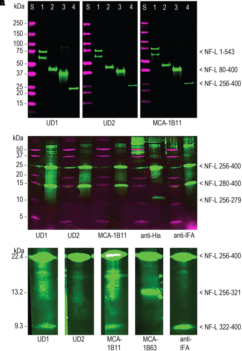
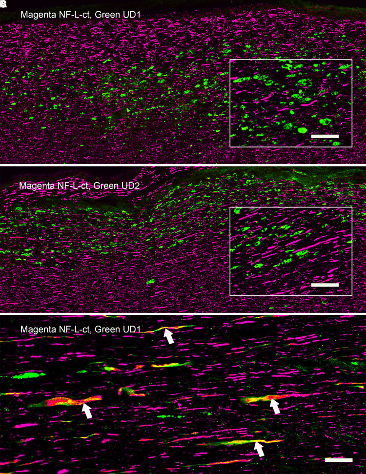
Recent work shows that certain antibody-based assays for the neurofilament light chain detect informative signals in the CSF and blood of human and animals affected by a variety of CNS injury and disease states. Much of this work has been performed using two mouse monoclonal antibodies to neurofilament light, UD1 and UD2, also known as Clones 2.1 and 47.3, respectively. These are the essential components of the Uman Diagnostics Neurofilament-Light™ ELISA kit, the Quanterix Simoa™ bead-based assay and others. We show that both antibodies bind to neighbouring epitopes in a short, conserved and unusual peptide in the centre of the neurofilament light Coil 2 segment of the 'rod' domain. We also describe a surprising and useful feature of Uman and similar reagents. While other well-characterized neurofilament antibodies generally show robust staining of countless cells and processes in CNS sections from healthy rats, both Uman antibodies reveal only a minor subset of profiles, presumably spontaneously degenerating or degenerated neurons and their processes. However, following experimental mid-cervical spinal cord injuries to rats, both Uman antibodies recognize numerous profiles in fibre tracts damaged by the injury administered. These profiles were typically swollen, beaded, discontinuous or sinusoidal as expected for degenerating and degenerated processes. We also found that several antibodies to the C-terminal 'tail' region of the neurofilament light protein bind undamaged axonal profiles but fail to recognize the Uman-positive material. The unmasking of the Uman epitopes and the loss of the neurofilament light tail epitopes can be mimicked by treating sections from healthy animals with proteases suggesting that the immunostaining changes we discovered are due to neurodegeneration-induced proteolysis. We have also generated a novel panel of monoclonal and polyclonal antibodies directed against the Uman epitopes that have degeneration-specific staining properties identical to the Uman reagents. Using these, we show that the region to which the Uman reagents bind contains further hidden epitopes distinct from those recognized by the two Uman reagents. We speculate that the Uman-type epitopes are part of a binding region important for higher order neurofilament assembly. The work provides important insights into the properties of the Uman assay, describes novel and useful properties of Uman-type and neurofilament light tail-binding antibodies and provides a hypothesis relevant to further understanding of neurofilament assembly.
Keywords: ELISA; NF-L; biomarker; neurodegeneration; neurofilament.
© The Author(s) 2023. Published by Oxford University Press on behalf of the Guarantors of Brain.
Conflict of interest statement
G.S. is founder and majority owner of EnCor Biotechnology Inc. and may gain income and equity from the sale of reagents described in this article. I.M., Y.L. and Y.W are full-time employees of EnCor Biotechnology and may also benefit from indirect income growth. S.R. and D.F. are full-time employees of the University of Florida and have no competing financial conflicts of interest.
Figures


References
-
- Stys PK. General mechanisms of axonal damage and its prevention. J Neurol Sci. 2005;233(1-2):3–13. - PubMed
-
- Buki A, Povlishock JT. All roads lead to disconnection?—Traumatic axonal injury revisited. Acta Neurochir (Wien). 2006; 148(2):181–193; discussion 193-4. - PubMed
-
- Shaw G, Yang C, Ellis R, et al. . Hyperphosphorylated neurofilament NF-H is a serum biomarker of axonal injury. Biochem Biophys Res Commun. 2005;336(4):1268–1277. - PubMed
-
- Shaw G. Chap 21 the use and potential of pNF-H as a general blood biomarker of axonal loss: An immediate application for CNS injury. In: Kobeissy FH, ed. Brain neurotrauma: Molecular, neuropsychological, and rehabilitation aspects. CRC Press, Taylar and Francis Group; 2015:289–300. - PubMed
LinkOut - more resources
Full Text Sources
Other Literature Sources