. 2023 Mar 30;5(2):fcad103.
doi: 10.1093/braincomms/fcad103. eCollection 2023.

Brain dysconnectivity with heart failure

Affiliations

Brain dysconnectivity with heart failure

Karsten Mueller et al. Brain Commun. .

Abstract

Structural brain damage associated with heart failure is well described; however, little is known about associated changes in various specific brain functions that bear immediate clinical relevance. A satisfactory pathophysiological link between heart failure and decline in cognitive function is still missing. In the present study, we aim to detect functional correlates of heart failure in terms of alterations in functional brain connectivity (quantified by functional magnetic resonance imaging) related to cognitive performance assessed by neuropsychological testing. Eighty patients were post hoc grouped into subjects with and without coronary artery disease. The coronary artery disease patients were further grouped as presenting with or without heart failure according to the guidelines of the European Society of Cardiology. On the basis of resting-state functional magnetic resonance imaging, brain connectivity was investigated using network centrality as well as seed-based correlation. Statistical analysis aimed at specifying centrality group differences and potential correlations between centrality and heart failure-related measures including left ventricular ejection fraction and serum concentrations of N-terminal fragment of the pro-hormone brain-type natriuretic peptide. The resulting correlation maps were then analysed using a flexible factorial model with the factors 'heart failure' and 'cognitive performance'. Our core findings are: (i) A statistically significant network centrality decrease was found to be associated with heart failure primarily in the precuneus, i.e. we show a positive correlation between centrality and left ventricular ejection fraction as well as a negative correlation between centrality and N-terminal fragment of the pro-hormone brain-type natriuretic peptide. (ii) Seed-based correlation analysis showed a significant interaction between heart failure and cognitive performance related to a significant decrease of precuneus connectivity to other brain regions. We obtained these results by different analysis approaches indicating the robustness of the findings we report here. Our results suggest that the precuneus is a brain region involved in connectivity decline in patients with heart failure, possibly primarily or already at an early stage. Current models of Alzheimer's disease-having pathophysiological risk factors in common with cerebrovascular disorders-also consider reduced precuneus connectivity as a marker of brain degeneration. Consequently, we propose that heart failure and Alzheimer's disease exhibit partly overlapping pathophysiological paths or have common endpoints associated with a more or less severe decrease in brain connectivity. This is further supported by specific functional connectivity alterations between the precuneus and widely distributed cortical regions, particularly in patients showing reduced cognitive performance.

Keywords: brain connectivity; cognitive impairment; functional magnetic resonance imaging; heart failure; precuneus.


Conflict of interest statement

The authors report no competing interests.

Figures

Graphical Abstract
Graphical Abstract
Figure 1
Figure 1
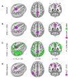
Brain network centrality decrease in patients with heart failure. Investigating global correlation (GCOR) of all three groups of patients with a full-factorial model including (i) patients with coronary artery disease (CAD) with heart failure (CAD+); (ii) CAD patients without heart failure (CAD−) and (iii) patients without CAD (no-CAD), we obtained a significant main effect using (A) an F-contrast across the group factor. Post hoc group comparisons were performed within the same model using t-contrasts and revealed a GCOR decrease (B) when comparing CAD+ with CAD− patients, and (C) when comparing CAD+ patients with no-CAD patients. All results were located in the precuneus but in no other brain region. Results were obtained with P < 0.05 using family-wise error (FWE) correction at cluster-level (see Table 1 for all comparisons including both centrality measures of GCOR and eigenvector centrality). Note, in a preceding study, a grey matter density (GMD) decrease was found with the same comparisons in the very same cohort particularly in the precuneus. (D) The obtained GCOR group differences are located in the same brain region within the precuneus, which was detected in a systemic and quantitative meta-analysis investigating patients converting from mild cognitive impairment to Alzheimer’s disease. ALE = activation likelihood estimation; L = left; R = right; x, y, z = coordinates in mm.
Figure 2
Figure 2
Brain network centrality decrease in patients with heart failure. A significant network centrality decrease was obtained when comparing patients with heart failure and coronary artery disease (CAD+) with all other patients without heart failure (ALL−). This network centrality decrease was obtained with both (A) global correlation (GCOR) and (B) eigenvector centrality (EC). With both GCOR and EC, centrality decrease was obtained in the precuneus but in no other brain region. Statistical analysis was performed using a general linear model with a full-factorial design implementing the group comparison using a t-contrast. Results were obtained with P < 0.05 using family-wise error (FWE) correction at cluster-level (see Table 1, bottom rows). L = left; R = right; x, y, z = coordinates in mm.
Figure 3
Figure 3
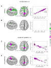
Correlation between heart failure-related biomarkers and brain network centrality across all patients. (A) A significant positive correlation was detected between the initial measurement of left ventricular ejection fraction (LVEF(1)) and global correlation (GCOR) in the precuneus. Reduced LVEF(1) values were associated with diminished GCOR. (B) The follow-up measurement LVEF(2) did not show any significant correlation with GCOR. (C) A significant negative correlation was found between the initial measurement of N-terminal fragment of brain natriuretic peptide (NT-proBNP(1)) and GCOR in the precuneus. (D) A significant negative correlation was also found between the follow-up measurement NT-proBNP(2) and GCOR in the same region, namely the precuneus. For both the initial and the follow-up measurements NT-proBNP(1) and NT-proBNP(2), respectively, higher serum levels were associated with diminished GCOR values. Statistical analyses were performed using a general linear model implementing a one-sample t-test including a t-contrast with the covariate of interest. Results were obtained with P < 0.05 using family-wise error (FWE) correction at cluster-level (see Tables 2 and 3). Note that, in a preceding study, a significant positive correlation was found between grey matter density (GMD) and both LVEF(1) and LVEF(2) in the same cohort of patients (see GMD in A and B). The same study shows also a negative correlation between GMD and NT-proBNP(1) (see GMD in C). L = left; M = image modality; R = right; x, y, z = coordinates in mm.
Figure 4
Figure 4
Brain connectivity decrease in patients showing a reduced cognitive performance with heart failure. (A) In patients with coronary artery disease and heart failure (CAD+), a seed-based connectivity (SBC) analysis revealed connectivity differences between patients with higher (HighCog) and lower (LowCog) cognitive performance. Diminished cognitive performance was found to be associated with a reduced brain connectivity between the precuneus and wide cortical areas including the motor system but also further frontal and parietal brain regions. (B) No SBC differences were found between HighCog and LowCog patients without heart failure (ALL−). (C) A significant interaction was obtained between the factors of heart failure (CAD+/ALL−) and cognition (HighCog/LowCog) showing diminished SBC with lower cognitive performance. Statistical analysis was performed using a general linear model with a full-factorial design implementing an interaction of both factors heart failure and cognitive performance. Significant results were obtained using a t-contrast with P < 0.05 using family-wise error (FWE) correction at cluster-level (see regions in magenta). L = left; R = right; x, y, z = coordinates in mm.

References

    1. Kalogeris T, Baines CP, Krenz M, Korthuis RJ. Cell biology of ischemia/reperfusion injury. Int Rev Cell Mol Biol. 2012;298:229–317. - PMC - PubMed
    1. Horstmann A, Frisch S, Jentzsch RT, Muller K, Villringer A, Schroeter ML. Resuscitating the heart but losing the brain: Brain atrophy in the aftermath of cardiac arrest. Neurology. 2010;74(4):306–312. - PubMed
    1. Mueller K, Thiel F, Beutner F, et al. . Brain damage with heart failure: Cardiac biomarker alterations and gray matter decline. Circ Res. 2020;126(6):750–764. - PMC - PubMed
    1. Ovsenik A, Podbregar M, Fabjan A. Cerebral blood flow impairment and cognitive decline in heart failure. Brain Behav. 2021;11(6):e02176. - PMC - PubMed
    1. Cannon JA, Moffitt P, Perez-Moreno AC, et al. . Cognitive impairment and heart failure: Systematic review and meta-analysis. J Card Fail. 2017;23(6):464–475. - PubMed

LinkOut - more resources