Optimum duration of hyperventilation during electroencephalography
- PMID: 37091767
- PMCID: PMC10120555
- DOI: 10.1080/08998280.2023.2177439
Optimum duration of hyperventilation during electroencephalography
Abstract
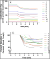
Hyperventilation (HV) is carried out for 3 minutes as a standard activation procedure in most routine electroencephalographic (EEG) procedures. The cerebral blood flow (CBF) reduction and the accompanying cerebral vasoconstriction caused by HV is believed to be the mechanism of EEG activation during HV. Some advocate for 5 minutes of HV, although the optimum duration is unknown. In this study, we measured the CBF continuously over the anterior temporal lobes using subdural probes, which use thermal diffusion flowmetry to measure CBF directly from the cerebral cortex. We sought to determine the duration of HV that produces the maximum reduction in CBF during routine HV in our epilepsy monitoring unit and prolonged the procedure for an additional 2 minutes for this study. Flowtronics® CBF probes were placed over the anterior temporal lobes in addition to the standard subdural strip placement for localization of their seizure focus in six patients who were candidates for epilepsy surgery. CBF was measured continuously for 2 minutes before and 5 minutes during HV for each patient. Time to reach maximum reduction of CBF for each attempt (11 temporal lobes) was computed. At 3 minutes, CBF reduction ranged from 11.6% to 40.0% from the pre-HV CBF level (mean 23.9%). At 5 minutes, CBF ranged from 14.3% to 42.0% (mean 25.7%). Six of the 11 measurements were steady or decreased slightly, and in the five other measurements, CBF showed a reverse trend after 3 minutes. A significant CBF reduction was attained in 3 minutes of HV in all trials. Continued HV after 3 minutes resulted in only a marginal (mean 1.8%) additional CBF reduction after 3 minutes. Thus, we propose that 3 minutes of HV is sufficient for EEG activation by the CBF criterion.
Keywords: Cerebral blood flow; electroencephalography; epilepsy; hyperventilation; seizure.
Copyright © 2023 Baylor University Medical Center.
Conflict of interest statement
The authors report no funding or conflicts of interest.
Figures


Similar articles
-
[Cerebral blood flow reactivity to hyperventilation in children with spontaneous occlusion of the circle of willis (moyamoya disease)].No Shinkei Geka. 1992 Apr;20(4):399-407. No Shinkei Geka. 1992. PMID: 1570062 Japanese.
-
Posthyperventilatory steal response in chronic cerebral hemodynamic stress: a positron emission tomography study.Stroke. 1998 Jul;29(7):1281-92. doi: 10.1161/01.str.29.7.1281. Stroke. 1998. PMID: 9660374
-
Diagnostic Yield of Five Minutes Compared to Three Minutes Hyperventilation During Electroencephalography in Children.Clin EEG Neurosci. 2023 Sep;54(5):522-525. doi: 10.1177/15500594211058266. Epub 2021 Nov 13. Clin EEG Neurosci. 2023. PMID: 34779251
-
Utility of daily supervised hyperventilation during long-term video-EEG monitoring.J Clin Neurophysiol. 2009 Feb;26(1):17-20. doi: 10.1097/WNP.0b013e3181969032. J Clin Neurophysiol. 2009. PMID: 19151614
-
Diagnostic yield of five minutes compared to three minutes hyperventilation during electroencephalography.Seizure. 2015 Aug;30:90-2. doi: 10.1016/j.seizure.2015.06.003. Epub 2015 Jun 14. Seizure. 2015. PMID: 26216691
References
-
- Foerster O, Penfield W.. The structural basis of traumatic epilepsy and results of radical operation. Brain. 1930;53(2):99–119. doi:10.1093/brain/53.2.99. - DOI
-
- Rosett J. The experimental production of rigidity, of abnormal involuntary movements and of abnormal states of consciousness in man. Brain. 1924;47(3):293–336. doi:10.1093/brain/47.3.293. - DOI
-
- Drury I. Electroencephalography. In Shah SM, Kelly KM, eds. Principles and Practice of Emergency Neurology. Cambridge, England: Cambridge University Press; 2003:30–34.
LinkOut - more resources
Full Text Sources
