

Genetic variations in the DYNC2H1 gene causing SRTD3 (short-rib thoracic dysplasia 3 with or without polydactyly)
- PMID: 37091781
- PMCID: PMC10116042
- DOI: 10.3389/fgene.2023.1125473
Genetic variations in the DYNC2H1 gene causing SRTD3 (short-rib thoracic dysplasia 3 with or without polydactyly)
Abstract
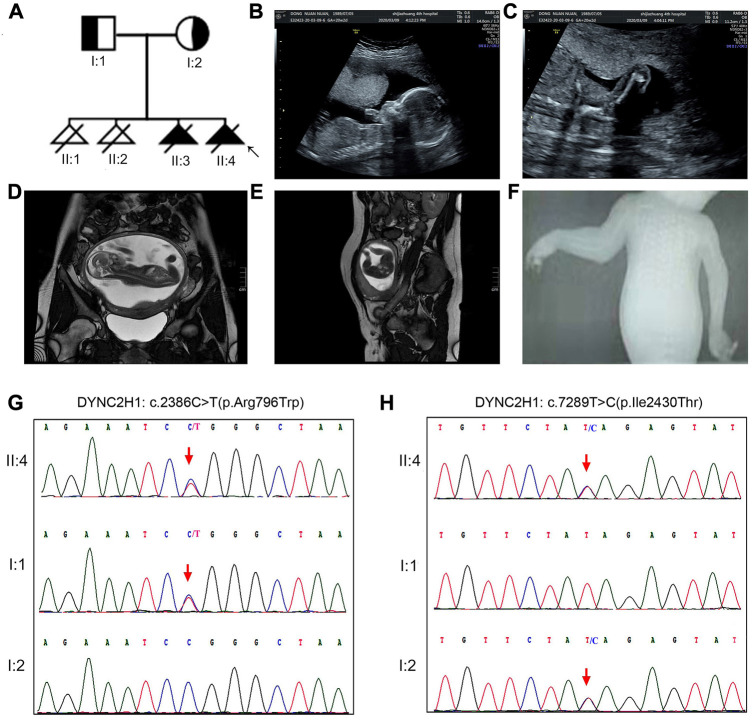
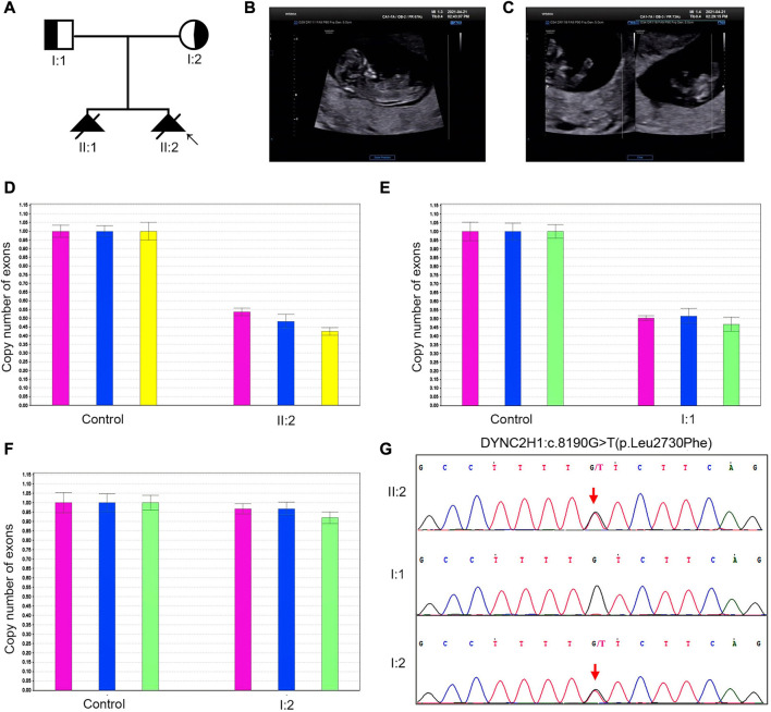
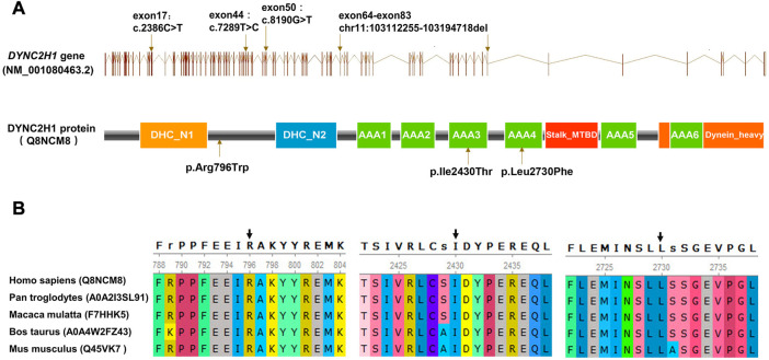
Background and aims: Short-rib thoracic dysplasia 3 with or without polydactyly (SRTD3) represents a type of severe fetal skeletal dysplasia (SD) characterized by shortened limbs, narrow thorax with or without polydactyly, which is caused by the homozygous or compound heterozygous mutations in the DYNC2H1 gene. SRTD3 is a recessive disorder, identification of the responsible genetic variation would be beneficial to an accurate prenatal diagnosis and well-grounded counseling for the affected families. Material and methods: Two families having experienced recurrent fetal SDs were recruited and submitted to a multiplatform genetic investigation. Whole-exome sequencing (WES) was performed with samples collected from the probands. Sanger sequencing and fluorescent quantitative PCR (qPCR) were conducted as validation assays for suspected variations. Results: WES identified two compound heterozygous variations in the DYNC2H1(NM_001080463.2) gene, namely c.2386C>T (p.Arg796Trp) and c.7289T>C (p.Ile2430Thr) for one; and exon (64-83)del and c.8190G>T (p.Leu2730Phe) for the other, respectively. One variant in them, exon (64-83)del, was novelly identified. Conclusion: The study detected two compound heterozygous variation in DYNC2H1 including one novel deletion: exon (64-83) del. Our findings clarified the cause of fetal skeletal dysplasia in the subject families, provided guidance for their future pregnancies, and highlighted the value of WES in diagnosis of skeletal dysplasia with unclear prenatal indications.
Keywords: DYNC2H1 gene; prenatal diagnosis; short-rib thoracic dysplasia 3 (SRTD3); skeletal dysplasia; whole-exome sequencing.
Copyright © 2023 Chen, Li, Zhang, Yuan, Sun, Yuan, Yang, Liang and Guo.
Conflict of interest statement
The authors declare that the research was conducted in the absence of any commercial or financial relationships that could be construed as a potential conflict of interest.
Figures
Similar articles
-
A novel compound heterozygous mutation in the DYNC2H1 gene in a Chinese family with Jeune syndrome.Hereditas. 2025 Jan 29;162(1):11. doi: 10.1186/s41065-025-00375-x. Hereditas. 2025. PMID: 39881416 Free PMC article.
-
Genetic analysis and prenatal diagnosis of short-rib thoracic dysplasia 3 with or without polydactyly caused by compound heterozygous variants of DYNC2H1 gene in four Chinese families.Front Genet. 2023 Mar 17;14:1075187. doi: 10.3389/fgene.2023.1075187. eCollection 2023. Front Genet. 2023. PMID: 37007936 Free PMC article.
-
Compound heterozygous variants in DYNC2H1 in a foetus with type III short rib-polydactyly syndrome and situs inversus totalis.BMC Med Genomics. 2022 Mar 12;15(1):55. doi: 10.1186/s12920-022-01205-z. BMC Med Genomics. 2022. PMID: 35277174 Free PMC article.
-
Prenatal diagnosis of short-rib polydactyly syndrome type III or short-rib thoracic dysplasia 3 with or without polydactyly (SRTD3) associated with compound heterozygous mutations in DYNC2H1 in a fetus.Taiwan J Obstet Gynecol. 2018 Feb;57(1):123-127. doi: 10.1016/j.tjog.2017.12.021. Taiwan J Obstet Gynecol. 2018. PMID: 29458881
-
Identification of novel compound heterozygous mutations of the DYNC2H1 gene in a fetus with short-rib thoracic dysplasia 3 with or without polydactyly.Intractable Rare Dis Res. 2020 May;9(2):95-98. doi: 10.5582/irdr.2020.01031. Intractable Rare Dis Res. 2020. PMID: 32494556 Free PMC article.
Cited by
-
Unclassifiable short-rib thoracic dysplasia diagnosed using targeted gene panel sequencing.Hum Genome Var. 2024 Dec 3;11(1):44. doi: 10.1038/s41439-024-00302-y. Hum Genome Var. 2024. PMID: 39622812 Free PMC article.
-
A novel compound heterozygous mutation in the DYNC2H1 gene in a Chinese family with Jeune syndrome.Hereditas. 2025 Jan 29;162(1):11. doi: 10.1186/s41065-025-00375-x. Hereditas. 2025. PMID: 39881416 Free PMC article.
-
Whole-Exome Sequencing Identifies DYNC2H1 Mutations as a Cause of Jeune Asphyxiating Thoracic Dystrophy Without Extra-Skeletal Organ Involvement.Int Med Case Rep J. 2024 Mar 23;17:209-214. doi: 10.2147/IMCRJ.S447466. eCollection 2024. Int Med Case Rep J. 2024. PMID: 38550721 Free PMC article.
References
LinkOut - more resources
Full Text Sources