. 2023 Apr 5:11:1170389.
doi: 10.3389/fcell.2023.1170389. eCollection 2023.

Absence of Nkx2-3 induces ectopic lymphatic endothelial differentiation associated with impaired extramedullary stress hematopoiesis in the spleen

Affiliations

Absence of Nkx2-3 induces ectopic lymphatic endothelial differentiation associated with impaired extramedullary stress hematopoiesis in the spleen

Fanni Gábris et al. Front Cell Dev Biol. .

Abstract

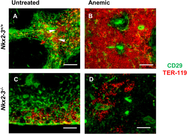
The red and white pulps as two main parts of the spleen are arranged around distinct types of vasculature, and perform significantly different functions in both humans and mice. Previous observations indicated a profound alteration of the local vessel specialization in mice lacking Nkx2-3 homeodomain transcription factor, including contradictory results suggesting presence of an ectopic lymphatic vascular structure. Furthermore, how the absence of Nkx2-3 and the consequential changes in endothelial components affect the extramedullary hematopoietic activity restricted to the splenic red pulp is unknown. In this work, we investigated the role of Nkx2-3 homeodomain transcription factor as a major morphogenic determinant for vascular specification, and its effect in the extramedullary hematopoiesis following acute blood loss and pharmacological stimulation of megakaryocyte differentiation after treatment with thrombopoietin-receptor mimetic Romiplostim. We found that, in mice lacking Nkx2-3, Prox1-positive lymphatic capillaries containing gp38/CD31 double positive lymphatic endothelial cells develop, arranged into an extensive meshwork, while the Clever1-positive venous segments of red pulp blood vasculature are absent. This lymphatic endothelial shift is coupled with a severely compromised splenic erythropoiesis and a significantly reduced splenic megakaryocyte colony formation following Romiplostim treatment in mice lacking Nkx2-3. These findings indicate that the shift of microvascular patterning in the absence of Nkx2-3 includes the emergence of ectopic Prox1-positive lymphatic vessels, and that this pivoting towards lymph node-like vascular patterning is associated with an impaired reserve hematopoietic capacity of the splenic red pulp.

Keywords: NKX2-3; Prox1; hematopoiesis; lymphatic endothelium; spleen; stroma.


Conflict of interest statement

The authors declare that the research was conducted in the absence of any commercial or financial relationships that could be construed as a potential conflict of interest.

Figures

FIGURE 1
FIGURE 1
Organziation of Prox1-expressing lymphatic capillaries in Nkx2-3−/− spleens. Compared to heterozygote sample (top) Nkx2-3-deficient mice have a reduced spleen size and patchy red pulp appearance (middle and bottom) (A). Following paraformaldehyde fixation and transparency treatment of the same samples, the heterozygote tissue (top) could not be clarified due to residual hemoglobin pigment, while the homozygote KO samples (middle and bottom) have become completely transparent (B). After CUBIC treatment in Prox1 GFP -Nkx2-3−/− mice (C) an extensive meshwork of LEC capillaries can be observed, while in Prox1 GFP -Nkx2-3+/- (D) such structures are absent, only faint background autofluorescence is detectable. Scale bars in A-B = 0.5 cm; scale bars in C-D = 100 μm. Representative images of a cohort of n = 3, experiment repeated twice.
FIGURE 2
FIGURE 2
Flow cytometric characterization of Prox1:eGFP+ cells in mice lacking Nkx2-3. Peripheral lymph nodes (pLN) were gated to include large stromal cells [(A), R1] and either eGFP [(B), R2/left] or eGFP+ [(B), R3/right] cells, then resolved according to their gp38(podoplanin)/CD31 features. Representative images demonstrate that the Prox1:eGFP+ (R3) cells in both pLN (D) and spleen (Spl, (F) from Nkx2-3−/− mice display gp38/CD31 double positive LEC phenotype (numbers indicating their frequency), whereas eGFP:Prox1- cells from Nkx2-3−/− pLN (C) and spleen (E) constitute non-LEC stromal subsets. In Nkx2-3+/- spleens no GFP+ signal is present (G). (H), gp38(podoplanin)/CD31 staining of GFP (R2) cells in Nkx2-3+/- spleens. Representative dot plots from n = 4, experiment repeated twice.
FIGURE 3
FIGURE 3
Nkx2-3 is necessary for the spleen red pulp vascular specification. Cryosections from C57Bl/6J mice were stained with the indicated antibodies, demonstrating co-localization of Clever1 (red) and IBL-9/2 (blue) staining (top) with relation to white pulp follicular dendritic meshwork (CR1/2, green) to visualize follicles (bottom, left) in wild-type spleen. Inset of the merged image demonstrates the vascular overlap between Clever1 and IBL-9/2 markers as purple (A). In wild-type mice (B) both Clever1 and IBL-9/2 labeling are present, whereas the absence of Nkx2-3 leads to the absence of both markers (C); in contrast, the deletion of Stab1 only abolishes Clever1 expression (D). Representative images, experiment repeated twice, n = 4, scale bar = 100 µm.
FIGURE 4
FIGURE 4
Hematopoiesis-supporting splenic stroma is disrupted in Nkx2-3−/− mice. Cryosections from Nkx2-3+/+ (A–D) and Nkx2-3−/− spleens (E–F) were stained for indicated markers, revealing intense expression of both CD29 and VCAM-1 (green) outside the white pulp delineated by CD45 staining (purple). Higher power images of the rectangles within the RP marked [in (C, G)] with dashed line demonstrate both stromal (arrows) and vascular (arrowheads) co-expression of CD29 and VCAM-1 [appearing as yellow, (D, H)]. Representative dual fluorescence microscopic images from a cohort of n = 4. Scale bars correspond to 100 μm.
FIGURE 5
FIGURE 5
Acute blood loss in Nkx2-3−/− mice induces imbalanced erythropoietic responses in the bonee marrow and spleen. Representative flow cytometric plots demonstrate results of the distribution of bone marrow and spleen lymphoid and erythroid cells in BALB/c and Nkx2-3−/− UTC and treated mice, numbers indicate relative frequency in the rectangles (A). Total number of lymphoid gated spleen cells in BALB/c (UTC n = 4, treated n = 4) and Nkx2-3−/− (UTC n = 3, treated n = 3) mice (B). Wet weight of spleen in BALB/c (UTC n = 4, treated n = 4) and Nkx2-3−/− (UTC n = 3, treated n = 3) mice (C). Ratio of splenic erythroid cells in BALB/c (UTC n = 10, treated n = 10) and Nkx2-3−/− (UTC n = 7, treated n = 8) mice (D). Absolute number of splenic erythroid cells in BALB/c (UTC n = 4, treated n = 4) and Nkx2-3−/− (UTC n = 3, treated n = 3) mice (E). Total number of lymphoid gated viable bone marrow cells in BALB/c (UTC n = 4, treated n = 4) and Nkx2-3−/− (UTC n = 3, treated n = 3) mice (F). Ratio of bone marrow erythroid cells in BALB/c (UTC n = 10, treated n = 10) and Nkx2-3−/− (UTC n = 7, treated n = 8) mice (G). Absolute number of bone marrow erythroid cells in BALB/c (UTC n = 4, treated n = 4) and Nkx2-3−/− (UTC n = 3, treated n = 3) mice (H). *p < 0.05, **p < 0.01, ***p < 0.001, ****p < 0.0001 by unpaired t-test and Mann Whitney U test.
FIGURE 6
FIGURE 6
Defective extramedullary expansion of splenic erythroid colonies in mice lacking Nkx2-3 compared to wild-type mice. Compared to untreated controls (A), in anemic BALB/c mice the TER-119-positive erythroid colonies (red, arrowheads) embedded in CD29-positive stroma (green) expand throughout the RP following blood withdrawal (B); in Nkx2-3−/− mice (C–D) only erythrocyte clusters are visible, without any noticeable change upon blood loss. Representative dual fluorescence microscopic images from a cohort of n = 4. Scale bars correspond to 100 μm.
FIGURE 7
FIGURE 7
TPO-agonist treatment fails to induce splenic megakaryocyte expansion in mice lacking Nkx2-3. Representative images show megakaryocyte labeling of BALB/c or Nkx2-3−/− (indicated at the top) spleen using anti-CD41 immunohistochemistry (visualized as brown reaction product, with hematoxylin counterstain) before (untreated, (A–C) and after Romiplostim treatment (D–F), indicated on the left. Higher power inserts (B, E) corresponding to the dashed rectangle demonstrate single megakaryocytes restricted to the RP. In the samples after Romiplostim treatment the arrows in the high-power insert (corresponding to the rectangle) label multinucleated megakaryocytes typically arranged in pairs. Representative micrographs from a cohort of n = 4. Scale bars correspond to 100 μm. Graphs demonstrate the number of CD41+ cells per 1 mm2 in BALB/c (UTC n = 3, treated n = 3) and Nkx2-3−/− (UTC n = 3, treated n = 3) mice (G). **p < 0.01 by unpaired t-test and Mann Whitney U test (G).
FIGURE 8
FIGURE 8
Sequence analysis of Prox1 promoter for putative binding sites for Nkx2-3 and COUP-TFII The transcription factors are indicated at the top, their binding sequence motifs are demonstrated according to JASPAR database, compared to the putative target sequences within the promoter and their sequence relative to transcription start site (TSS).

Similar articles

Cited by

References

    1. Alexandre Y. O., Schienstock D., Lee H. J., Gandolfo L. C., Williams C. G., Devi S., et al. (2022). A diverse fibroblastic stromal cell landscape in the spleen directs tissue homeostasis and immunity. Sci. Immunol. 7, eabj0641. 10.1126/sciimmunol.abj0641 - DOI - PubMed
    1. Balázs M., Horváth G., Grama L., Balogh P. (2001). Phenotypic identification and development of distinct microvascular compartments in the postnatal mouse spleen. Cell. Immunol. 212, 126–137. 10.1006/cimm.2001.1847 - DOI - PubMed
    1. Balogh P., Balázs M., Czömpöly T., Weih D. S., Arnold H. H., Weih F. (2007). Distinct roles of lymphotoxin-beta signaling and the homeodomain transcription factor Nkx2.3 in the ontogeny of endothelial compartments in spleen. Cell. Tissue Res. 328, 473–486. 10.1007/s00441-007-0378-6 - DOI - PubMed
    1. Bellomo A., Mondor I., Spinelli L., Lagueyrie M., Stewart B. J., Brouilly N., et al. (2020). Reticular fibroblasts expressing the transcription factor WT1 define a stromal niche that maintains and replenishes splenic red pulp macrophages. Immunity 53, 127–142.e7. 10.1016/j.immuni.2020.06.008 - DOI - PubMed
    1. Bellomo A., Gentek R., Golub R., Bajénoff M. (2021). Macrophage-fibroblast circuits in the spleen. Immunol. Rev. 302, 104–125. 10.1111/imr.12979 - DOI - PubMed

LinkOut - more resources