

Accuracy of the newly developed Zimmer Biomet Root Aiming guide in tibial tunnel creation compared with that of conventional guides
- PMID: 37092123
- PMCID: PMC10120359
- DOI: 10.1016/j.asmart.2023.03.001
Accuracy of the newly developed Zimmer Biomet Root Aiming guide in tibial tunnel creation compared with that of conventional guides
Abstract
Background/objective: Accurate tibial tunnel creation is crucial for successful transtibial pullout repair of medial meniscus (MM) posterior root tears (MMPRTs). This study aimed to evaluate the accuracy of the newly developed Zimmer Biomet Root Aiming (ZeBRA) guide for transtibial pullout repair of MMPRTs.
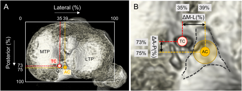
Methods: This study included 50 patients who underwent transtibial pullout repair using the Unicorn Meniscal Root (UMR) (n = 25) and ZeBRA (n = 25) guides. The expected anatomic centre (AC) and tibial tunnel centre (TC) were assessed using three-dimensional postoperative computed tomography (CT) images. The expected AC was defined as the centre of the circle tangent to the triangular footprint of the MM posterior root. The expected AC and TC on the tibial surface were assessed using the percentage-based posterolateral location on the tibial surface. The absolute distance between the AC and TC (mm) was evaluated.
Results: The mean AC location was 76.1% ± 3.1% posterior and 40.8% ± 2.1% lateral, whereas the mean TC location was 76.7% ± 5.3% posterior and 37.2% ± 3.6% lateral using the UMR guide and 75.8% ± 3.1% posterior and 36.5% ± 2.4% lateral using the ZeBRA guide. No significant difference was observed in the absolute distance between the UMR and ZeBRA guides (3.9 ± 1.4 and 3.8 ± 1.3 mm, respectively; p = 0.617).
Conclusions: The newly developed ZeBRA guide allows accurate tibial tunnel creation, and its accuracy is comparable to that of the conventional UMR guide. Tibial tunnels were created at optimal positions using both guides, and the choice of the guide would depend on the surgeon's preference.
Keywords: Meniscus; Musculoskeletal diseases; Orthopaedic procedures; Tibial tunnel; Zimmer biomet root aiming guide.
© 2023 Asia Pacific Knee, Arthroscopy and Sports Medicine Society. Published by Elsevier (Singapore) Pte.
Conflict of interest statement
The authors have no conflicts of interest to declare relevant to this article.
Figures


Similar articles
-
A newly-developed guide can create tibial tunnel at an optimal position during medial meniscus posterior root repairs.J Orthop Sci. 2022 Jul;27(4):815-820. doi: 10.1016/j.jos.2021.04.002. Epub 2021 May 24. J Orthop Sci. 2022. PMID: 34039522
-
The accuracy of a newly developed guide system in medial meniscus posterior root repair: a comparison between two aiming guides.Knee Surg Relat Res. 2019 Aug 7;31(1):7. doi: 10.1186/s43019-019-0007-1. Knee Surg Relat Res. 2019. PMID: 32660577 Free PMC article.
-
A new aiming guide can create the tibial tunnel at favorable position in transtibial pullout repair for the medial meniscus posterior root tear.Orthop Traumatol Surg Res. 2017 May;103(3):367-371. doi: 10.1016/j.otsr.2017.01.005. Epub 2017 Feb 24. Orthop Traumatol Surg Res. 2017. PMID: 28238962
-
Arthroscopic transtibial pullout repair for posterior meniscus root tears.Oper Orthop Traumatol. 2019 Jun;31(3):248-260. doi: 10.1007/s00064-018-0574-4. Epub 2018 Oct 26. Oper Orthop Traumatol. 2019. PMID: 30367186 Review. English.
-
Posterior Root Meniscal Tears: Preoperative, Intraoperative, and Postoperative Imaging for Transtibial Pullout Repair.Radiographics. 2016 Oct;36(6):1792-1806. doi: 10.1148/rg.2016160026. Radiographics. 2016. PMID: 27726749 Review.
Cited by
-
Minimizing the risk of injury to the popliteal artery during pullout repair of medial meniscus posterior root tears: A cadaveric study.Asia Pac J Sports Med Arthrosc Rehabil Technol. 2024 Jan 17;35:81-84. doi: 10.1016/j.asmart.2023.11.009. eCollection 2024 Jan. Asia Pac J Sports Med Arthrosc Rehabil Technol. 2024. PMID: 38261907 Free PMC article.
-
Diagnosis and Treatment Strategies of Meniscus Root Tears: A Scoping Review.Orthop J Sports Med. 2024 Nov 1;12(11):23259671241283962. doi: 10.1177/23259671241283962. eCollection 2024 Nov. Orthop J Sports Med. 2024. PMID: 39493310 Free PMC article.
References
-
- LaPrade C.M., James E.W., Cram T.R., Feagin J.A., Engebretsen L., LaPrade R.F. Meniscal root tears: a classification system based on tear morphology. Am J Sports Med. 2015;43:363–369. - PubMed
-
- Furumatsu T., Okazaki Y., Okazaki Y., et al. Injury patterns of medial meniscus posterior root tears. Orthop Traumatol Surg Res. 2019;105:107–111. - PubMed
-
- Krych A.J., Reardon P.J., Johnson N.R., et al. Non-operative management of medial meniscus posterior horn root tears is associated with worsening arthritis and poor clinical outcome at 5-year follow-up. Knee Surg Sports Traumatol Arthrosc. 2017;25:383–389. - PubMed
-
- Allaire R., Muriuki M., Gilbertson L., Harner C.D. Biomechanical consequences of a tear of the posterior root of the medial meniscus. Similar to total meniscectomy. J Bone Joint Surg Am. 2008;90:1922–1931. - PubMed
-
- LaPrade C.M., Foad A., Smith S.D., et al. Biomechanical consequences of a nonanatomic posterior medial meniscal root repair. Am J Sports Med. 2015;43:912–920. - PubMed
LinkOut - more resources
Full Text Sources
