

Structural-Functional Correlates of Response to Pedunculopontine Stimulation in a Randomized Clinical Trial for Axial Symptoms of Parkinson's Disease
- PMID: 37092235
- PMCID: PMC10357146
- DOI: 10.3233/JPD-225031
Structural-Functional Correlates of Response to Pedunculopontine Stimulation in a Randomized Clinical Trial for Axial Symptoms of Parkinson's Disease
Abstract
Background: Axial symptoms of Parkinson's disease (PD) can be debilitating and are often refractory to conventional therapies such as dopamine replacement therapy and deep brain stimulation (DBS) of the subthalamic nuclei (STN).
Objective: Evaluate the efficacy of bilateral DBS of the pedunculopontine nucleus area (PPNa) and investigate structural and physiological correlates of clinical response.
Methods: A randomized, double-blind, cross-over clinical trial was employed to evaluate the efficacy of bilateral PPNa-DBS on axial symptoms. Lead positions and neuronal activity were evaluated with respect to clinical response. Connectomic cortical activation profiles were generated based on the volumes of tissue activated.
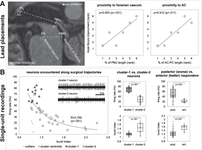
Results: PPNa-DBS modestly improved (p = 0.057) axial symptoms in the medication-off condition, with greatest positive effects on gait symptoms (p = 0.027). Electrode placements towards the anterior commissure (ρ= 0.912; p = 0.011) or foramen caecum (ρ= 0.853; p = 0.031), near the 50% mark of the ponto-mesencephalic junction, yielded better therapeutic responses. Recording trajectories of patients with better therapeutic responses (i.e., more anterior electrode placements) had neurons with lower firing-rates (p = 0.003) and higher burst indexes (p = 0.007). Structural connectomic profiles implicated activation of fibers of the posterior parietal lobule which is involved in orienting behavior and locomotion.
Conclusion: Bilateral PPNa-DBS influenced gait symptoms in patients with PD. Anatomical and physiological information may aid in localization of a favorable stimulation target.
Keywords: Deep brain stimulation; Parkinson’s disease; pedunculopontine nucleus.
Conflict of interest statement
S.B. has ownership interest in Neurostar GmbH (not related to this work). A.G. was supported by research grants (not related to this work) from Medtronic, Boston Scientific, Abbott, the Baden-Wuerttemberg Foundation, and the German Federal Ministry of Education and Research. D.W. has received research support from the German Research Council and the Michael J. Fox Foundation (not related to this work). L.M. has received honoraria from Medtronic (nor related to this work). G.N., I.C. have no conflicts of interest to report.
Figures


Similar articles
-
Bilateral deep brain stimulation of the pedunculopontine and subthalamic nuclei in severe Parkinson's disease.Brain. 2007 Jun;130(Pt 6):1596-607. doi: 10.1093/brain/awl346. Epub 2007 Jan 24. Brain. 2007. PMID: 17251240
-
Deep brain stimulation of the pedunculopontine nucleus modulates subthalamic pathological oscillations.Neurobiol Dis. 2019 Aug;128:49-52. doi: 10.1016/j.nbd.2018.11.006. Epub 2018 Nov 10. Neurobiol Dis. 2019. PMID: 30423476
-
Combined STN/SNr-DBS for the treatment of refractory gait disturbances in Parkinson's disease: study protocol for a randomized controlled trial.Trials. 2011 Oct 11;12:222. doi: 10.1186/1745-6215-12-222. Trials. 2011. PMID: 21989388 Free PMC article. Clinical Trial.
-
Pedunculopontine nucleus deep brain stimulation in Parkinson's disease: A clinical review.Mov Disord. 2018 Jan;33(1):10-20. doi: 10.1002/mds.27098. Epub 2017 Sep 28. Mov Disord. 2018. PMID: 28960543 Review.
-
Effects of deep brain stimulation on balance and gait in patients with Parkinson's disease: A systematic neurophysiological review.Neurophysiol Clin. 2015 Nov;45(4-5):371-88. doi: 10.1016/j.neucli.2015.07.001. Epub 2015 Aug 28. Neurophysiol Clin. 2015. PMID: 26319759 Review.
Cited by
-
A Pragmatic Review on Spinal Cord Stimulation Therapy for Parkinson's Disease Gait Related Disorders: Gaps and Controversies.Mov Disord Clin Pract. 2024 Aug;11(8):927-947. doi: 10.1002/mdc3.14143. Epub 2024 Jun 20. Mov Disord Clin Pract. 2024. PMID: 38899557 Free PMC article.
-
Nigral stimulation for freezing of gait: kinematic gait parameters inform optimization of stimulation frequency.J Neuroeng Rehabil. 2025 Sep 9;22(1):191. doi: 10.1186/s12984-025-01712-x. J Neuroeng Rehabil. 2025. PMID: 40926219 Free PMC article. Clinical Trial.
-
The Middle Managers: Thalamic and Cholinergic Contributions To Coordinating Top-Down And Bottom-Up Processing.Curr Opin Behav Sci. 2024 Aug;58:101406. doi: 10.1016/j.cobeha.2024.101406. Epub 2024 May 16. Curr Opin Behav Sci. 2024. PMID: 39220566 Free PMC article.
-
Vestibular Neurostimulation for Parkinson's Disease: A Novel Device-Aided Non-Invasive Therapeutic Option.J Pers Med. 2024 Aug 31;14(9):933. doi: 10.3390/jpm14090933. J Pers Med. 2024. PMID: 39338187 Free PMC article. Review.
-
Hypo-connectivity of the primary somatosensory cortex in Parkinson's disease: a resting-state functional MRI study.Front Neurol. 2024 Apr 30;15:1361063. doi: 10.3389/fneur.2024.1361063. eCollection 2024. Front Neurol. 2024. PMID: 38746656 Free PMC article.
References
-
- Ferraye MU, Debû B, Fraix V, Xie-Brustolin J, Chabardès S, Krack P, Benabid A-L, Pollak P (2008) Effects of subthalamic nucleus stimulation and levodopa on freezing of gait in Parkinson disease. Neurology 70, 1431. - PubMed
-
- Breit S, Bouali-Benazzouz R, Benabid A-L, Benazzouz A (2001) Unilateral lesion of the nigrostriatal pathway induces an increase of neuronal activity of the pedunculopontine nucleus, which is reversed by the lesion of the subthalamic nucleus in the rat. Eur J Neurosci 14, 1833–1842. - PubMed
-
- Rinne JO, Ma SY, Lee MS, Collan Y, Röyttä M (2008) Loss of cholinergic neurons in the pedunculopontine nucleus in Parkinson’s disease is related to disability of the patients. Parkinsonism Relat Disord 14, 553–557. - PubMed
-
- Karachi C, Grabli D, Bernard FA, Tandé D, Wattiez N, Belaid H, Bardinet E, Prigent A, Nothacker H-P, Hunot S, Hartmann A, Lehéricy S, Hirsch EC, Francois C (2010) Cholinergicmesencephalic neurons are involved in gait and postural disorders inParkinson disease. J Clin Invest 120, 2745–2754. - PMC - PubMed
Publication types
MeSH terms
LinkOut - more resources
Full Text Sources
Medical
