. 2023 Apr 24;8(8):e164178.
doi: 10.1172/jci.insight.164178.

MKRN3 inhibits puberty onset via interaction with IGF2BP1 and regulation of hypothalamic plasticity

Affiliations

MKRN3 inhibits puberty onset via interaction with IGF2BP1 and regulation of hypothalamic plasticity

Lydie Naulé et al. JCI Insight. .

Abstract

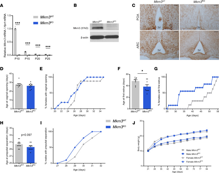
Makorin ring finger protein 3 (MKRN3) was identified as an inhibitor of puberty initiation with the report of loss-of-function mutations in association with central precocious puberty. Consistent with this inhibitory role, a prepubertal decrease in Mkrn3 expression was observed in the mouse hypothalamus. Here, we investigated the mechanisms of action of MKRN3 in the central regulation of puberty onset. We showed that MKRN3 deletion in hypothalamic neurons derived from human induced pluripotent stem cells was associated with significant changes in expression of genes controlling hypothalamic development and plasticity. Mkrn3 deletion in a mouse model led to early puberty onset in female mice. We found that Mkrn3 deletion increased the number of dendritic spines in the arcuate nucleus but did not alter the morphology of GnRH neurons during postnatal development. In addition, we identified neurokinin B (NKB) as an Mkrn3 target. Using proteomics, we identified insulin-like growth factor 2 mRNA-binding protein 1 (IGF2BP1) as another target of MKRN3. Interactome analysis revealed that IGF2BP1 interacted with MKRN3, along with several members of the polyadenylate-binding protein family. Our data show that one of the mechanisms by which MKRN3 inhibits pubertal initiation is through regulation of prepubertal hypothalamic development and plasticity, as well as through effects on NKB and IGF2BP1.

Keywords: Endocrinology; Mouse models; Neurodevelopment; Neuroendocrine regulation; Neuroscience.


Figures

Figure 1
Figure 1. Generation of MKRN3-WT and MKRN3-knockout hiPSC-derived hypothalamic ARC neurons.
(A) Schematic representation of the differentiation protocol for the generation of hypothalamic neurons from MKRN3-WT and MKRN3-KO hiPSCs, including live-cell imaging of hiPSCs before differentiation, neural progenitors (NPCs) on day 16 of differentiation, and hypothalamic ARC neurons on day 30 of differentiation. (BI) MKRN3 (B), OCT4 (C), NKX2.1 (D), NESTIN (E), MAP2 (F), POMC (G), KISS1 (H), and TAC3 (I) mRNA levels (relative to levels in MKRN3-WT 1 NPCs) in hiPSCs, NPCs, and hypothalamic ARC neurons derived from the MKRN3-WT 1 and MKRN3-KO 1 clones (n = 3 differentiation protocols per group). Data are presented as the mean ± SEM. Statistics were performed using 2-way ANOVA, followed by Tukey’s post hoc test. *P < 0.01. KO, knockout; OCT4, octamer-binding transcription factor 4; NKX2.1, NK2 homeobox 1; NESTIN, Nestin; MAP2, microtubule associated protein 2; POMC, proopiomelanocortin.
Figure 2
Figure 2. Comparison of transcriptomes of MKRN3-KO and MKRN3-WT hiPSC-derived hypothalamic ARC neurons reveals differences in expression of genes that control hypothalamic neuronal development and plasticity.
(A) Volcano plot comparing Benjamini-Hochberg–adjusted (BH-adjusted) P values against fold-change, showing transcripts that are differentially expressed between MKRN3-KO and MKRN3-WT hypothalamic ARC neurons (genes downregulated in MKRN3-KO neurons in blue, genes upregulated in MKRN3-KO neurons in orange, and genes not significantly different in gray). The analysis was performed using a BH adjusted P value cutoff of 0.05 and a log2 fold-change ratio cutoff of 1. Three representative genes more highly expressed in MKRN3-KO are shown. SLIT1, slit guidance ligand 1; SLIT2, slit guidance ligand 2; NRCAM, neuronal cell adhesion molecule. (B) Heatmap of the top 50 genes that are the most differentially expressed in MKRN3-KO 1 compared with MKRN3-WT 1 hypothalamic neurons. Each column represents 1 differentiation protocol. (C) Dot plot of Gene Ontology (GO) terms enriched between MKRN3-WT and MKRN3-KO hypothalamic neurons. The size of the dots represents the number of genes in the GO term, and the color gradient indicates the adjusted P value, using the BH method. (DF) Heatmaps of differentially expressed genes (DEGs) in 3 significantly enriched pathways: (D) extracellular matrix (ECM) organization, (E) axon guidance, and (F) synapse organization, in MKRN3-KO compared with MKRN3-WT hypothalamic neurons. (G) Heatmap for a subset of DEGs between MKRN3-WT and MKRN3-KO hypothalamic neurons that have been associated with age at menarche in GWAS. (H) Relative SLIT1, SLIT2, and NRCAM mRNA levels in MKRN3-WT 1, MKRN3-WT 2, MKRN3-KO 1, and MKRN3-KO 2 hypothalamic neurons (n = 3 differentiation protocols per group). Data are presented as the mean ± SEM values. Statistics were performed using 2-way ANOVA, followed by Tukey’s post hoc test. *P < 0.05, **P < 0.01.
Figure 3
Figure 3. Mkrn3 deletion in mice is associated with early onset of puberty in female mice and a tendency toward early puberty in male mice.
(A) Relative Mkrn3 mRNA levels in the ARC of Mkrn3WT and Mkrn3KO females across PND10, PND15, PND20, and PND25 (n = 6 per genotype and age). Statistics were performed using 2-way ANOVA, followed by Tukey’s post hoc test. (B) Representative Western blot autoradiographic image of Mkrn3 protein and β-actin in the MBH of Mkrn3WT and Mkrn3KO females at PND10. (C) Representative immunohistochemistry images of Mkrn3 protein in the POA and the arcuate nucleus (ARC) of Mkrn3WT and Mkrn3KO females at PND10. Scale bar = 100 μm. 3V, third ventricle. (DG) Age at vaginal opening (D) and cumulative percentage of female mice exhibiting vaginal opening (E), and age at first estrus (F) and cumulative percentage of female mice with first estrus (G), in Mkrn3WT (n = 8) and Mkrn3KO (n = 10) females. (H) Age at preputial separation and (I) cumulative percentage of male mice exhibiting preputial separation in Mkrn3WT (n = 10) and Mkrn3KO (n = 11) males. Statistics were performed using unpaired 2-tailed t test. (J) Body weight of Mkrn3WT (n = 11) and Mkrn3KO (n = 15) male and Mkrn3WT (n = 8) and Mkrn3KO (n = 10) female mice measured weekly from weaning (PND21) to adulthood (PND84). Statistics were performed using 2-way ANOVA, followed by Tukey’s post hoc test. Data are presented as the mean ± SEM values. *P < 0.05, ***P < 0.0001 of Mkrn3KO compared with Mkrn3WT.
Figure 4
Figure 4. Mkrn3 deletion in mice does not affect postnatal hypothalamic expression of Kiss1, Tac3, Tac1, Gnrh1, or Kiss1r.
(AC) Relative mRNA levels of (A) Kiss1, (B) Tac3, and (C) Tac1 in the ARC of Mkrn3WT and Mkrn3KO females across postnatal development (PND10, PND15, PND20, and PND25) (n = 6 per genotype and per age). (DF) Relative mRNA levels of (D) Kiss1, (E) Gnrh1, and (F) Kiss1r in the POA of Mkrn3WT and Mkrn3KO females at PND10, PND15, PND20, and PND25 (n = 6 per genotype and per age). Statistics were performed using 2-way ANOVA, followed by Tukey’s post hoc test. Data are presented as the mean ± SEM. Bars with different letters are significantly different from one another (P < 0.05).
Figure 5
Figure 5. Mkrn3 deletion in mice increases NKB protein levels in the ARC.
(AD) Representative images of (A) GnRH-immunoreactive (GnRH-ir) neurons in the rPOA and (B) fiber immunoreactivity in the ME of Mkrn3WT and Mkrn3KO females at PND10. (C) Quantification of the number of GnRH-ir neurons in the rPOA and (D) mean density of GnRH immunoreactivity in the ME (n = 5 per genotype). (EH) Representative images of kisspeptin immunoreactivity in the periventricular nuclei (PeN) of the RP3V of Mkrn3WT and Mkrn3KO females at (E) PND15 and (F) PND25. Quantification of the number of kisspeptin-ir cells in the anteroventral periventricular (AVPV) area and the PeN at (G) PND15 and (H) PND25 (n = 5–6 per age and per genotype). (IL) Representative images of kisspeptin immunoreactivity in the ARC of Mkrn3WT and Mkrn3KO females at (I) PND15 and (J) PND25. Quantification of the mean density of kisspeptin immunoreactivity in the ARC at (K) PND15 and (L) PND25 (n = 6 per age and genotype). (MP) Representative images of NKB immunoreactivity in the ARC of Mkrn3WT and Mkrn3KO females at (M) PND15 and (N) PND25. Quantification of the mean density of NKB immunoreactivity in the ARC at (O) PND15 and (P) PND25 (n = 5–6 per age and genotype). Scale bar = 100 μm. Statistics were performed using unpaired 2-tailed t test. Data are presented as the mean ± SEM values. *P < 0.05 compared with Mkrn3WT.
Figure 6
Figure 6. Mkrn3 deletion does not alter GnRH neuron morphology but increases spine density in the ARC.
(AC) Representative images of GnRH neurons classified into (A) unipolar, (B) bipolar, and (C) complex dendritic morphology. Scale bar = 50 μm. (D) Quantification of the percentage of GnRH neurons in the rPOA with unipolar, bipolar, and complex morphology in Mkrn3WT and Mkrn3KO females at PND15 (n = 5 per genotype). Statistics were performed using 2-way ANOVA, followed by Tukey’s post hoc test. Data are presented as median and distribution of the data and probability density (violin plot). (E) Representative images of Golgi-Cox–impregnated neurons in the ARC of Mkrn3WT and Mkrn3KO females at PND15. Scale bar = 5 μm. (F) Quantification of the number of dendritic spines/50 μm in ARC neurons of Mkrn3WT and Mkrn3KO females at PND15 (n = 5 per genotype), classified as thin, stubby, or mushroom according to their morphology. Statistical analysis was performed using unpaired 2-tailed t tests. Data are presented as median and distribution of the data and probability density (violin plot). *P < 0.05 compared with Mkrn3WT.
Figure 7
Figure 7. Mkrn3 deletion increases Igf2bp1 protein levels in the ARC.
(A) Volcano plot illustrating the significant changes in abundance of detected peptides in Mkrn3KO compared with Mkrn3WT mice in the ARC at PND15. Vertical lines represent a 25% increase in fold-change; horizontal line represents an FDR of 0.01 for Mkrn3WT (n = 2) and Mkrn3KO (n = 3) male and Mkrn3WT (n = 3) and Mkrn3KO (n = 3) female mice. (B and C) Relative TMT signal-to-noise levels for (B) Mkrn3 and (C) Igf2bp1 proteins in the ARC of PND15 Mkrn3WT (n = 2) and Mkrn3KO (n = 3) male and Mkrn3WT (n = 3) and Mkrn3KO (n = 3) female mice. TMT RA, tandem mass tag relative abundance. (D) Representative images of Igf2bp1 immunoreactivity in the ARC of Mkrn3WT and Mkrn3KO females at PND15. (E) Quantification of the mean density of Igf2bp1 immunoreactivity in the ARC of Mkrn3WT (n = 6) and Mkrn3KO (n = 5) females at PND15. Scale bar = 100 μm. Statistics were performed using unpaired 2-tailed t test. Data are presented as mean ± SEM. *P < 0.05 compared with Mkrn3WT mice.
Figure 8
Figure 8. MKRN3 interacts with IGF2BP1.
(A) Interactome map of key protein interactions with MKRN3. The network includes MKRN3 detected by different purification methods. Circle sizes indicate the CompPASS interaction score. Green circles indicate interaction identified in HEK293 cells, pink circles in SH-SY5Y cells, and orange circles in both cell lines. The oval nodes represent different clusters of prey proteins. (B) Co-IP analysis of MKRN3 and IGF2BP1 interaction. HEK293T cells were transiently transfected with HA-MKRN3, GFP-IGF2BP1, or both. Lysates were immunoprecipitated using anti-HA antibody. Both input and co-IP fractions were immunoblotted using anti-IGF2BP1 or anti-HA antibodies. The immunoblot demonstrates that IGF2BP1 is co-immunoprecipitated by anti-HA antibodies when coexpressed with HA-MKRN3.

References

    1. Abreu AP et al. Central precocious puberty caused by mutations in the imprinted gene MKRN3. N Engl J Med. 2013;368(26):2467–2475. doi: 10.1056/NEJMoa1302160. - DOI - PMC - PubMed
    1. Maione L, et al. Makorin RING finger protein 3 and central precocious puberty. Curr Opin Endocr Metab Res. 2020;14:152–159. doi: 10.1016/j.coemr.2020.08.003. - DOI - PMC - PubMed
    1. Valadares LP, et al. MKRN3 mutations in central precocious puberty: a systematic review and meta-analysis. J Endocr Soc. 2019;3(5):979–995. doi: 10.1210/js.2019-00041. - DOI - PMC - PubMed
    1. Jong MT, et al. A novel imprinted gene, encoding a RING zinc-finger protein, and overlapping antisense transcript in the Prader-Willi syndrome critical region. Hum Mol Genet. 1999;8(5):783–793. doi: 10.1093/hmg/8.5.783. - DOI - PubMed
    1. Jong MT, et al. Imprinting of a RING zinc-finger encoding gene in the mouse chromosome region homologous to the Prader-Willi syndrome genetic region. Hum Mol Genet. 1999;8(5):795–803. doi: 10.1093/hmg/8.5.795. - DOI - PubMed

Publication types