. 2023 Jun;10(17):e2206521.
doi: 10.1002/advs.202206521. Epub 2023 Apr 24.

Lymph Node Follicle-Targeting STING Agonist Nanoshells Enable Single-Shot M2e Vaccination for Broad and Durable Influenza Protection

Affiliations

Lymph Node Follicle-Targeting STING Agonist Nanoshells Enable Single-Shot M2e Vaccination for Broad and Durable Influenza Protection

Hsiao-Han Tsai et al. Adv Sci (Weinh). 2023 Jun.

Abstract

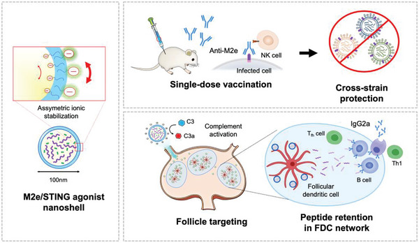
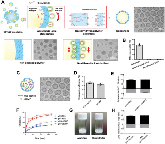
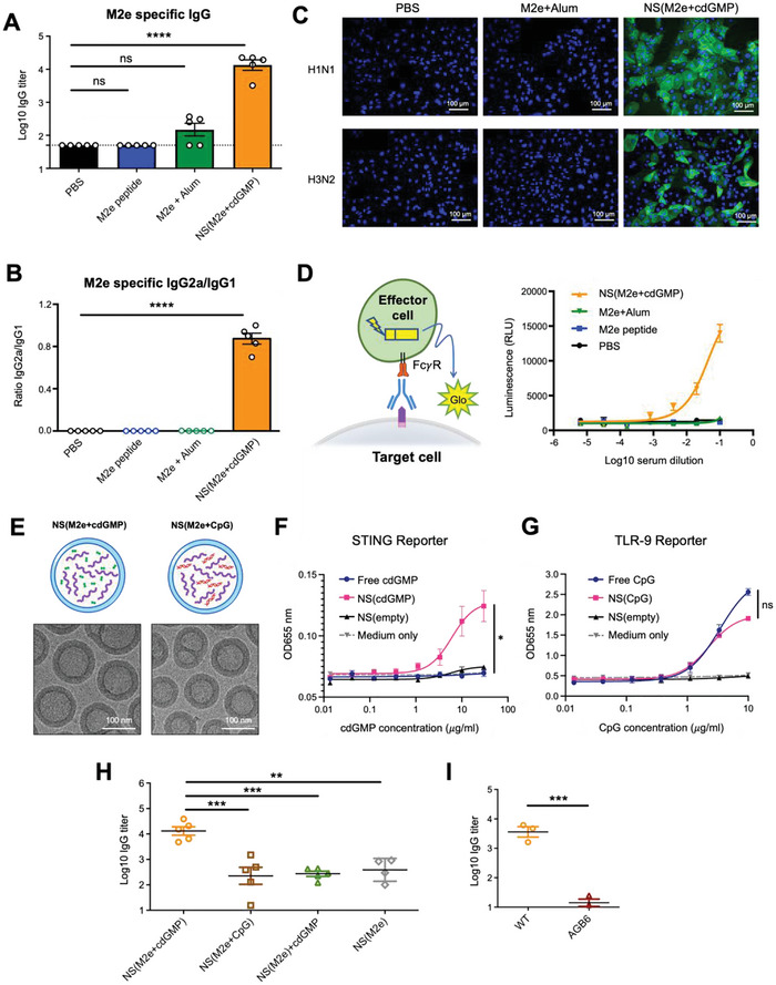
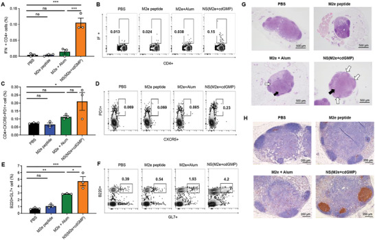
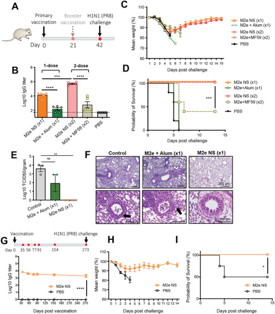
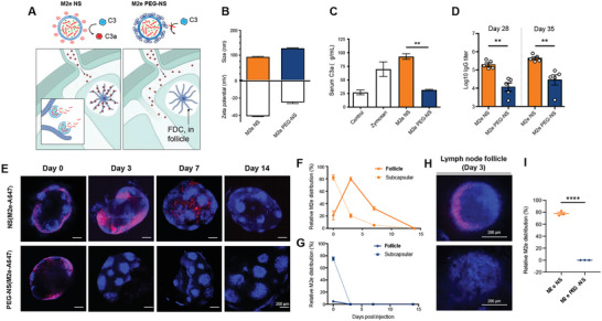
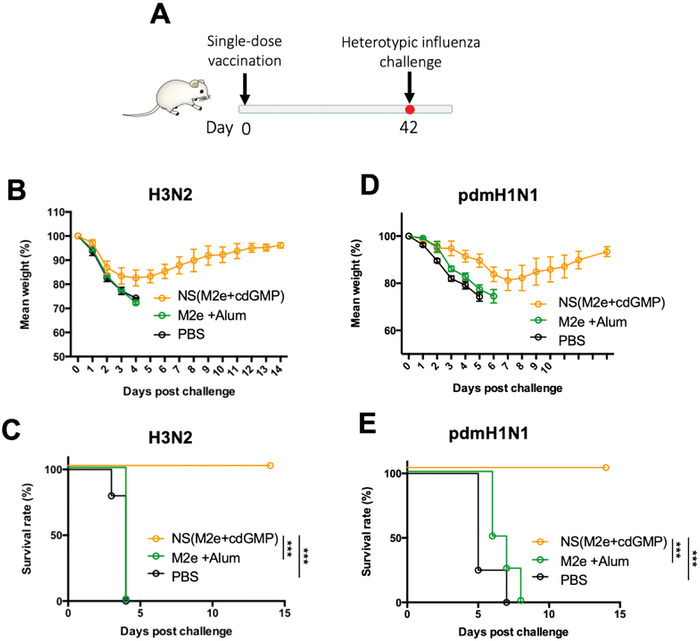
The highly conserved matrix protein 2 ectodomain (M2e) of influenza viruses presents a compelling vaccine antigen candidate for stemming the pandemic threat of the mutation-prone pathogen, yet the low immunogenicity of the diminutive M2e peptide renders vaccine development challenging. A highly potent M2e nanoshell vaccine that confers broad and durable influenza protectivity under a single vaccination is shown. Prepared via asymmetric ionic stabilization for nanoscopic curvature formation, polymeric nanoshells co-encapsulating high densities of M2e peptides and stimulator of interferon genes (STING) agonists are prepared. Robust and long-lasting protectivity against heterotypic influenza viruses is achieved with a single administration of the M2e nanoshells in mice. Mechanistically, molecular adjuvancy by the STING agonist and nanoshell-mediated prolongation of M2e antigen exposure in the lymph node follicles synergistically contribute to the heightened anti-M2e humoral responses. STING agonist-triggered T cell helper functions and extended residence of M2e peptides in the follicular dendritic cell network provide a favorable microenvironment that induces Th1-biased antibody production against the diminutive antigen. These findings highlight a versatile nanoparticulate design that leverages innate immune pathways for enhancing the immunogenicity of weak immunogens. The single-shot nanovaccine further provides a translationally viable platform for pandemic preparedness.

Keywords: follicular dendritic cells; germinal center; lymph node follicle targeting; matrix protein 2 ectodomain antigen; nanoshell; stimulator of interferon genes agonist; universal influenza vaccine.


Conflict of interest statement

The authors declare no conflict of interest.

Figures

Figure 1
Figure 1
Design (left), application (top right), and mechanism (bottom right) of a single‐shot M2e‐based influenza vaccine for broad influenza protection.
Figure 2
Figure 2
Preparation and characterization of M2e nanoshell vaccine. A) Schematics for the asymmetrically stabilized nanoemulsion process for nanoshell vaccine preparation and cryoEM images of polymeric nanoshells. The absence of charged polymer or differential ionic buffers led to emulsion collapse. B) Quantification of M2e peptide encapsulation following different emulsion processes for nanoparticle preparation. C) CryoEM visualization of M2e nanoshell vaccine co‐encapsulating M2e peptide antigens and cdGMP. Scale bars = 100 nm. D) Encapsulation efficiency of M2e peptides and cdGMP by the M2e nanoshell vaccine. E) The size and zeta potential of empty nanoshells (NS(empty)) and M2e nanoshell vaccine (NS(M2e+cdGMP)) were measured by dynamic light scattering. F) Release kinetics of M2e peptides and cdGMP from nanoshell vaccines at pH 5 and pH 7. G) Images of M2e nanoshell vaccine following lyophilization and reconstitution. H) The size and zeta potential of M2e nanoshells as measured by DLS show comparable physicochemical properties before and after lyophilization.
Figure 3
Figure 3
Anti‐M2e induction and ADCC activity following M2e nanoshell vaccine inoculation in mice. A) M2e‐specific IgG titers following a single shot immunization with PBS, M2e peptide, Alum‐adjuvanted M2e peptides, and M2e nanoshell vaccine in mice. B) M2e‐specific IgG2a to IgG1 titer ratios in immunized Balb/C mice on day 35 post‐vaccination. Error bars represent mean ± SEM (N = 5). C) Acetone‐fixed MDCK cells with heterotypic influenza infections for evaluating antibody binding to cell‐bound M2e by anti‐M2e from mice serum. Immunofluorescence assays were performed using mice serum derived on day 42. Nuclei were stained with DAPI. H1N1: A/Puerto Rico/8/1934 (H1N1); H3N2: A/HKx31 (H3N2). Scale bars = 100 µm. D) ADCC surrogate assay with mice serum derived on day 42 post‐vaccination against H1N1‐infected MDCK cells. Data are presented as mean ± SEM. (N = 3). E) CryoEM images showing the morphology of nanoshells encapsulating the combinations of either M2e + cdGMP or M2e + CpG‐ODN 1826. F) Assessment of human STING activation by SEAP reporter cells with free cdGMP, NS(cdGMP), or empty NS following incubation for 24 h. G) Assessment of human TLR9 activation by SEAP reporter cells with free CpG‐ODN2395, NS(CpG‐ODN2395), or empty NS following incubation for 24 h. H) M2e‐specific IgG antibodies in BALB/C mice immunized with NS(M2e+cdGMP), NS(M2e+CpG), NS(M2e) + free cdGMP or NS(M2e) via the subcutaneous route. Error bars represent mean ± SEM (N = 5). I) M2e‐specific IgG antibodies in C57BL/6 mice or AGB6 mice immunized with NS(M2e+cdGMP). Error bars represent mean ± SEM (N = 3). Statistical analyses were performed by one‐way ANOVA or Student's t‐test (*p < 0.05, **p < 0.01, ***p < 0.001, ****p < 0.0001).
Figure 4
Figure 4
M2e STING agonist nanoshell promotes a lymph node environment favorable to Th1‐skewed antibody production. A,B) M2e‐specific IFNγ +CD4+ T cell responses in immunized mice as determined by intracellular cytokine staining on day 7 following primary immunization. Error bars represent mean ± SEM (N = 3). C,D) Frequencies of and E,F) GL7+ germinal center B cells in the draining lymph nodes of immunized mice 14 days after immunization. Error bars represent mean ± SEM (N = 3). Statistical analyses were performed by one‐way ANOVA (*p < 0.05, **p < 0.01, ***p < 0.001). G) Perfused popliteal lymph nodes of immunized mice 14 days post‐vaccination were fixed and embedded in paraffin. Haematoxylin/eosin staining (H&E) was performed to identify follicular hyperplasia (white arrows) and paracortex hyperplasia (black arrows) in the lymph nodes. Scale bars = 500 µm. H) Popliteal lymph node sections were stained with anti‐GL‐7 antibodies (brown) for germinal center identification. Scale bars = 200 µm.
Figure 5
Figure 5
Assessment of antiviral protectivity by single‐shot M2e nanoshell vaccination. A) Vaccination schedule and viral challenge for single‐shot and prime‐boost immunization regimens. B) Assessment of anti‐M2e titers from mice serum collected on day 35 following the primary vaccination. C) Mouse body weight changes and D) survival rate after A/Puerto Rico/8/1934 (H1N1) infection upon challenge on day 42 with 3 × 10⁵ PFU viral dose via the intranasal route. (N = 5). E) Lung viral titers were evaluated 3 days following virus infection. (N = 3). F) Haematoxylin/eosin staining (H&E) was performed to identify lymphocytic cell infiltrates and perivascular inflammation (top panel). Scale bars = 200 µm. The lung tissues were also monitored for bronchiole injuries (bottom panel), including the presence of necrotic epithelial cells (white arrows) and airway wall thickening (black arrows). Scale bars = 100 µm. G) Anti‐M2e titers in mice following a primary M2e nanoshell vaccination over a 273‐day period. (N = 5). H) Mice were challenged on day 273 intranasally with 3 × 10⁵ PFU of A/Puerto Rico/8/1934 (H1N1) and assessed for (H) body weight changes and I) survival rate after infection. (N = 5). Error bars represent mean ± SEM. Statistical analyses were performed by one‐way ANOVA. The survival rate was analyzed by using the Log‐rank test (**p < 0.01, ***p < 0.001, ****p < 0.0001; ns, non‐significant).
Figure 6
Figure 6
Spatiotemporal control of M2e peptide antigen distribution in the lymph node follicle by nanoshell carriers. A) Schematics illustrating the effect of nanoshell surface property on complement activation and lymph node distribution. B) Dynamic light scattering characterization of size and zeta potential of PEG‐free M2e STING agonist nanoshells (M2e NS) and PEG‐coated M2e STING agonist nanoshells (M2e PEG‐NS). (N = 3). C) The activated complement protein C3a concentration in BALB/c mouse serum (Control) and following incubation with zymosan (Zymosan), PEG‐free nanoshells (M2e NS), or PEGylated nanoshells (M2e PEG‐NS) (N = 3). D) M2e‐specific IgG titers in mice 35 days following immunization with M2e NS or M2e PEG‐NS. Error bars represent mean ± SEM (N = 5). Statistical analyses were performed by unpaired t‐tests. (**p < 0.01). E) BALB/c mice were inoculated with nanoshells containing fluorescent A647‐conjugated M2e peptide (M2e‐A647) for tracking of M2e antigen distribution over a 14‐day period. FDCs were labeled in situ with anti‐CD35 antibody and excised dLNs were cleared and imaged by confocal microscopy (CD35 blue; M2e‐A647 red). Scale bars = 200 µm. F,G) Co‐localization of M2e with subcapsular macrophages and lymph node follicles was evaluated via image analysis of M2e‐A647 signal coordination with lymph node boundaries and anti‐CD35 signals, respectively. H) Zoomed‐in visualization of M2e distribution in the lymph node follicles three days following subcutaneous administrated with either M2e NS or M2e PEG‐NS. Scale bars = 200 µm. I) Quantification of M2e NS and M2e PEG‐NS in the lymph node follicles 3 days following inoculation. (N = 3) (****p < 0.0001).
Figure 7
Figure 7
Assessment of M2e nanoshell vaccine against heterotypic influenza challenge. A) Vaccination and viral challenge schedule. Mice were subcutaneously inoculated with PBS, M2e NS, or free M2e peptides adjuvanted with Alum. The mice were challenged on day 42 intranasally with 3 × 10⁶ PFU influenza A/HKx31 (H3N2) or 2.5 × 10⁶ PFU pandemic 2009 H1N1 (pdmH1N1). B) Mouse body weight changes and C) survival rate after influenza A/HKx31 (H3N2) infection. D) Mouse body weight changes and E) survival rate after the influenza pandemic 2009 H1N1 (pdmH1N1) infection. Error bars represent mean ± SEM. (N = 5). The survival rate was analyzed by using the Log‐rank test (*** p < 0.001).

Similar articles

Cited by

References

    1. Gordon A., Reingold A., Curr. Epidemiol. Rep 2018, 5, 1. - PMC - PubMed
    1. a) Petrova V. N., Russell C. A., Nat. Rev. Microbiol. 2018, 16, 60; - PubMed
    2. b) Keshavarz M., Mirzaei H., Salemi M., Momeni F., Mousavi M. J., Sadeghalvad M., Arjeini Y., Solaymani‐Mohammadi F., Nahand J. S., Namdari H., Mokhtari‐Azad T., Rezaei F., Rev. Med. Virol. 2019, 29, e2014. - PubMed
    1. Subbarao K., Murphy B. R., Fauci A. S., Immunity 2006, 24, 5. - PubMed
    1. Belongia E. A., Kieke B. A., Donahue J. G., Greenlee R. T., Balish A., Foust A., Lindstrom S., Shay D. K., J. Infect. Dis. 2009, 199, 159. - PubMed
    1. a) Nachbagauer R., Feser J., Naficy A., Bernstein D. I., Guptill J., Walter E. B., Berlanda‐Scorza F., Stadlbauer D., Wilson P. C., Aydillo T., Behzadi M. A., Bhavsar D., Bliss C., Capuano C., Carreno J. M., Chromikova V., Claeys C., Coughlan L., Freyn A. W., Gast C., Javier A., Jiang K., Mariottini C., McMahon M., McNeal M., Solorzano A., Strohmeier S., Sun W., Van der Wielen M., Innis B. L., et al., Nat. Med. 2021, 27, 106; - PubMed
    2. b) Darricarrere N., Qiu Y., Kanekiyo M., Creanga A., Gillespie R. A., Moin S. M., Saleh J., Sancho J., Chou T. H., Zhou Y. F., Zhang R. J., Dai S. J., Moody A., Saunders K. O., Crank M. C., Mascola J. R., Graham B. S., Wei C. J., Nabel G. J., Sci. Transl. Med. 2021, 13, eabe5449; - PubMed
    3. c) Tseng Y. C., Wu C. Y., Liu M. L., Chen T. H., Chiang W. L., Yu Y. H., Jan J. T., Lin K. I., Wong C. H., Ma C., Proc. Natl. Acad. Sci. USA 2019, 116, 4200; - PMC - PubMed
    4. d) Laursen N. S., Friesen R. H. E., Zhu X., Jongeneelen M., Blokland S., Vermond J., van Eijgen A., Tang C., van Diepen H., Obmolova G., van der Neut Kolfschoten M., Zuijdgeest D., Straetemans R., Hoffman R. M. B., Nieusma T., Pallesen J., Turner H. L., Bernard S. M., Ward A. B., Luo J., Poon L. L. M., Tretiakova A. P., Wilson J. M., Limberis M. P., Vogels R., Brandenburg B., Kolkman J. A., Wilson I. A., Science 2018, 362, 598; - PMC - PubMed
    5. e) Lin P. H., Liang C. Y., Yao B. Y., Chen H. W., Pan C. F., Wu L. L., Lin Y. H., Hsu Y. S., Liu Y. H., Chen P. J., Hu C. J., Yang H. C., Mol. Ther. Methods Clin. Dev. 2021, 21, 299; - PMC - PubMed
    6. f) Boyoglu‐Barnum S., Ellis D., Gillespie R. A., Hutchinson G. B., Park Y. J., Moin S. M., Acton O. J., Ravichandran R., Murphy M., Pettie D., Matheson N., Carter L., Creanga A., Watson M. J., Kephart S., Ataca S., Vaile J. R., Ueda G., Crank M. C., Stewart L., Lee K. K., Guttman M., Baker D., Mascola J. R., Veesler D., Graham B. S., King N. P., Kanekiyo M., Nature 2021, 592, 623. - PMC - PubMed

Publication types