. 2023 May 1;79(Pt 5):409-419.
doi: 10.1107/S2059798323002322. Epub 2023 Apr 24.

Conformational transition of the Ixodes ricinus salivary serpin Iripin-4

Affiliations

Conformational transition of the Ixodes ricinus salivary serpin Iripin-4

Barbora Kascakova et al. Acta Crystallogr D Struct Biol. .

Abstract

Iripin-4, one of the many salivary serpins from Ixodes ricinus ticks with an as-yet unexplained function, crystallized in two different structural conformations, namely the native partially relaxed state and the cleaved serpin. The native structure was solved at a resolution of 2.3 Å and the structure of the cleaved conformation was solved at 2.0 Å resolution. Furthermore, structural changes were observed when the reactive-centre loop transitioned from the native conformation to the cleaved conformation. In addition to this finding, it was confirmed that Glu341 represents a primary substrate-recognition site for the inhibitory mechanism. The presence of glutamate instead of the typical arginine in the P1 recognition site of all structurally characterized I. ricinus serpins (PDB entries 7b2t, 7pmu and 7ahp), except for the tyrosine in the P1 site of Iripin-2 (formerly IRS-2; PDB entry 3nda), would explain the absence of inhibition of the tested proteases that cleave their substrate after arginine. Further research on Iripin-4 should focus on functional analysis of this interesting serpin.

Keywords: Iripin-4; Ixodes ricinus; X-ray structure; cleaved conformation; native conformation; serpins.


Figures

Figure 1
Figure 1
Analysis of heat-stable complex formation between Iripin-4 and tested proteases. No complex formation was observed. There is visible cleavage of Iripin-4 by porcine elastase, as shown by a black arrow. Proteins were resolved on 12% NuPAGE Bis-Tris gels and visualized by Coomassie staining.
Figure 2
Figure 2
The relationship between Iripin-4 concentration and k obs, the observed second-order rate constant for the binding of human granzyme B to Iripin-4. k obs was determined using a para-nitroanilide substrate (Ac-IEPD-pNA).
Figure 3
Figure 3
The RCL of native Iripin-4. (a) Front view of the RCL (blue) showing the inserted residues into β-sheet A (cyan) and highly flexible residues of the hinge region of the RCL (purple). (b) View rotated by 50°.
Figure 4
Figure 4
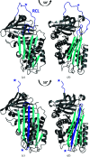
Crystal structures of Iripin-4. (a) Native Iripin-4; the position of the RCL partially inserted into the breach region of β-sheet A is highlighted. (b) Side view of the native conformation. (c) Cleaved Iripin-4 displaying an additional β-strand in β-sheet A as a result of RCL insertion. (d) Side view of the cleaved conformation. Both crystal structures are displayed as cartoons; β-sheet A is in cyan and the RCL is in blue. The position of the P1 cleavage site is marked by a blue asterisk.
Figure 5
Figure 5
Structural superposition of Iripin-4 conformations. The native Iripin-4 (cyan) with an exposed RCL (blue) is aligned with cleaved Iripin-4 (grey) containing a new additional β-strand (marine) labelled s4A. The three regions responsible for insertion of the RCL into β-sheet A are marked in circles (Khan et al., 2011 ▸). The hinge region is marked with an oval.
Figure 6
Figure 6
Superposition and alignment of I. ricinus native serpin conformations. (a) Superposition of the Iripin-4 (PDB entry 7zbf), Iripin-1 (PDB entry 7qtz; Chlastáková et al., 2023 ▸) and Iripin-8 (PDB entry 7pmu) crystal structures displayed as cartoons. The structure models are differentiated as follows: Iripin-4 is in blue, Iripin-1 is in red and Iripin-8 is in green. The structure of Iripin-1 was modified because of the absence of residues in its RCL, which are coloured salmon for emphasis. (b) A closer view of the RCL residues displayed as sticks; the colours are as described previously. (c) Structure-based sequence alignment of I. ricinus native serpins. The secondary-structure elements are depicted as arrows for β-strands and spirals for α-helices. The RCL is highlighted in a black box and the P1 and P1′ residues are marked according to the nomenclature of Schechter & Berger (1967 ▸).
Figure 7
Figure 7
Superposition and alignment of I. ricinus cleaved serpin conformations. (a) Superposition of the Iripin-4 (PDB entry 7zas), Iripin-2 (PDB entry 3nda), Iripin-3 (PDB entry 7ahp) and Iripin-5 (PDB entry 7b2t) crystal structures displayed as cartoons. The structure models are differentiated as follows: Iripin-4 is in blue, Iripin-2 is in grey, Iripin-3 is in magenta and Iripin-5 is in cyan. The RCL of all serpins is coloured salmon. (b) Structure-based sequence alignment of I. ricinus cleaved serpins. The secondary-structure elements are marked with arrows for β-strands and spirals for α-helices. The inserted RCL is highlighted in the black box and the P1 and P1′ residues are marked according to the nomenclature of Schechter & Berger (1967 ▸).

Similar articles

Cited by

  • Genome sequences of four Ixodes species expands understanding of tick evolution.
    Cerqueira de Araujo A, Noel B, Bretaudeau A, Labadie K, Boudet M, Tadrent N, Istace B, Kritli S, Cruaud C, Olaso R, Deleuze JF, Voordouw MJ, Hervet C, Plantard O, Zamoto-Niikura A, Chertemps T, Maïbèche M, Hilliou F, Le Goff G, Chmelař J, Mazák V, Jmel MA, Kotsyfakis M, Medina JM, Hackenberg M, Šimo L, Koutroumpa FA, Wincker P, Kopáček P, Perner J, Aury JM, Rispe C. Cerqueira de Araujo A, et al. BMC Biol. 2025 Jan 21;23(1):17. doi: 10.1186/s12915-025-02121-1. BMC Biol. 2025. PMID: 39838418 Free PMC article.

References

    1. Boulanger, N., Boyer, P., Talagrand-Reboul, E. & Hansmann, Y. (2019). Med. Mal. Infect. 49, 87–97. - PubMed
    1. Chen, V. B., Arendall, W. B., Headd, J. J., Keedy, D. A., Immormino, R. M., Kapral, G. J., Murray, L. W., Richardson, J. S. & Richardson, D. C. (2010). Acta Cryst. D66, 12–21. - PMC - PubMed
    1. Chlastáková, A., Kaščáková, B., Kotál, J., Langhansová, H., Kotsyfakis, M., Kutá Smatanová, I., Tirloni, L. & Chmelař, J. (2023). Front. Immunol. 14, 1116324. - PMC - PubMed
    1. Chlastáková, A., Kotál, J., Beránková, Z., Kaščáková, B., Martins, L. A., Langhansová, H., Prudnikova, T., Ederová, M., Kutá Smatanová, I., Kotsyfakis, M. & Chmelař, J. (2021). Front. Immunol. 12, 626200. - PMC - PubMed
    1. Chmelar, J., Oliveira, C. J., Rezacova, P., Francischetti, I. M. B., Kovarova, Z., Pejler, G., Kopacek, P., Ribeiro, J. M. C., Mares, M., Kopecky, J. & Kotsyfakis, M. (2011). Blood, 117, 736–744. - PMC - PubMed