. 2023 May 31;97(5):e0003023.
doi: 10.1128/jvi.00030-23. Epub 2023 Apr 24.

Specific Residues in the C-Terminal Domain of the Human Metapneumovirus Phosphoprotein Are Indispensable for Formation of Viral Replication Centers and Regulation of the Function of the Viral Polymerase Complex

Affiliations

Specific Residues in the C-Terminal Domain of the Human Metapneumovirus Phosphoprotein Are Indispensable for Formation of Viral Replication Centers and Regulation of the Function of the Viral Polymerase Complex

Rachel Erin Thompson et al. J Virol. .

Abstract

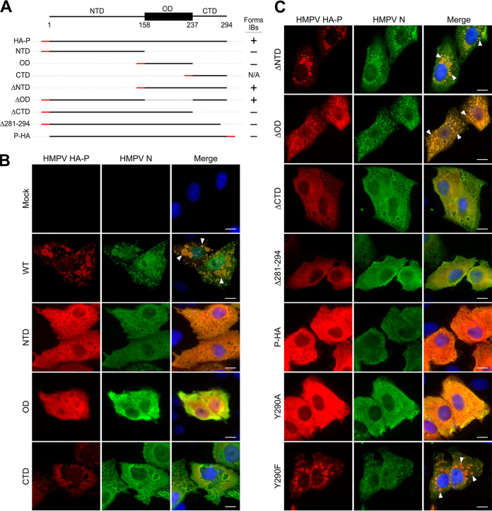
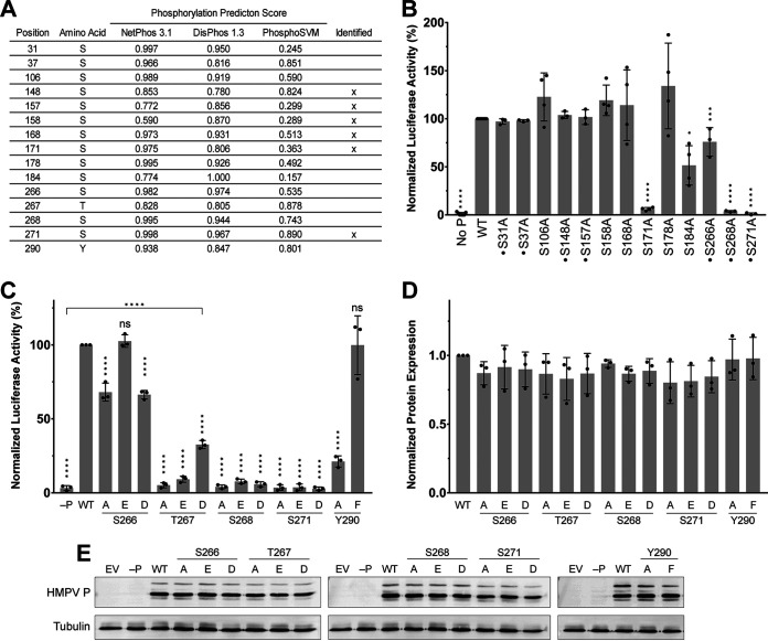
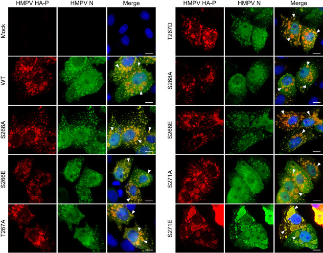
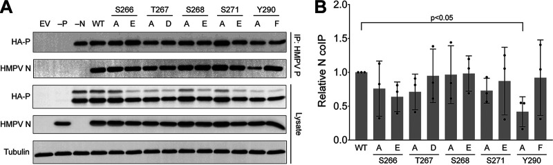
Human metapneumovirus (HMPV) is a negative-strand RNA virus that frequently causes respiratory tract infections in infants, the elderly, and the immunocompromised. A hallmark of HMPV infection is the formation of membraneless, liquid-like replication and transcription centers in the cytosol termed inclusion bodies (IBs). The HMPV phosphoprotein (P) and nucleoprotein (N) are the minimal viral proteins necessary to form IB-like structures, and both proteins are required for the viral polymerase to synthesize RNA during infection. HMPV P is a homotetramer with regions of intrinsic disorder and has several known and predicted phosphorylation sites of unknown function. In this study, we found that the P C-terminal intrinsically disordered domain (CTD) must be present to facilitate IB formation with HMPV N, while either the N-terminal intrinsically disordered domain or the central oligomerization domain was dispensable. Alanine substitution at a single tyrosine residue within the CTD abrogated IB formation and reduced coimmunoprecipitation with HMPV N. Mutations to C-terminal phosphorylation sites revealed a potential role for phosphorylation in regulating RNA synthesis and P binding partners within IBs. Phosphorylation mutations which reduced RNA synthesis in a reporter assay produced comparable results in a recombinant viral rescue system, measured as an inability to produce infectious viral particles with genomes containing these single P mutations. This work highlights the critical role HMPV P plays in facilitating a key step of the viral life cycle and reveals the potential role for phosphorylation in regulating the function of this significant viral protein. IMPORTANCE Human metapneumovirus (HMPV) infects global populations, with severe respiratory tract infections occurring in infants, the elderly, and the immunocompromised. There are currently no FDA-approved therapeutics available to prevent or treat HMPV infection. Therefore, understanding how HMPV replicates is vital for the identification of novel targets for therapeutic development. During HMPV infection, viral RNA synthesis proteins localize to membraneless structures called inclusion bodies (IBs), which are sites of genome replication and transcription. The HMPV phosphoprotein (P) is necessary for IBs to form and for the virus to synthesize RNA, but it is not known how this protein contributes to IB formation or if it is capable of regulating viral replication. We show that the C-terminal domain of P is the location of a molecular interaction driving IB formation and contains potential phosphorylation sites where amino acid charge regulates the function of the viral polymerase complex.

Keywords: inclusion bodies; phosphoprotein; pneumovirus; protein phosphorylation; replication complex.


Conflict of interest statement

The authors declare no conflict of interest.

Figures

FIG 1
FIG 1
Domains of HMPV P involved in IB formation. (A) Schematic of HMPV P domains (top: NTD, N-terminal domain; OD, oligomerization domain; CTD, C-terminal domain), HA-tagged domain constructs (HA tag location represented by a red line), and the constructs’ ability to colocalize with HMPV N to form IBs (right column). (B and C) HMPV HA-P WT or the HA-tagged P construct was cotransfected with HMPV N in Vero cells. Cells were labeled with antibodies for anti-HA (detecting P [red]) and anti-N (green), and nuclei were stained with DAPI (blue). IB formation was determined through fluorescence microscopy with a Nikon Ti2 inverted microscope with a 100× objective. Examples of IBs are indicated by white arrowheads. Scale bar represents 10 μm. Representative images were selected from triplicate experiments.
FIG 2
FIG 2
Effect of phosphorylation site mutagenesis on HMPV P function. (A) Summary of investigated P phosphorylation sites. The sequence of P from strain CAN97-83 was analyzed using phosphorylation prediction sites. Prediction scores for selected residues are listed on a 0-to-1 scale, and phosphorylated sites that have been identified (24) are indicated in the last column. (B) BSR T7/5 cells were transfected with an HMPV minireplicon system with WT HA-P or HA-P serine-to-alanine mutants and assayed for expression of the luciferase reporter as a measure of luminescence. (C) The minireplicon assay was repeated using phospho-dead and phosphomimetic mutations of C-terminal phosphorylation sites. P mutant expression was confirmed through quantification (D) of Western blot analysis (E). All experiments were completed in at least triplicate, and mutant values were normalized to the WT. Statistical analysis for panels B to D was completed using one-way ANOVA with Dunnett’s multiple-comparison test. *, P < 0.05; **, P < 0.005; ***, P < 0.0005; ****, P < 0.0001. Error bars represent 1 standard deviation.
FIG 3
FIG 3
IB formation of HMPV HA-P phosphorylation site mutants. Vero cells were cotransfected with HMPV N and HMPV HA-P WT or HA-P mutant. IBs were detected using antibodies for anti-HA (red) and anti-N (green), and nuclei were stained using DAPI (blue). IB formation was determined through fluorescence microscopy with a Nikon Ti2 inverted microscope with a 100× objective. Examples of IBs are indicated by white arrowheads. Scale bar represents 10 μm. Representative images were selected from duplicate experiments.
FIG 4
FIG 4
Coimmunoprecipitation of N and HA-P mutants. (A) HEK 293T cells were cotransfected with HMPV N and HA-P construct. HA-P WT and mutants were immunoprecipitated using Dynabeads chemically coupled to a nanobody specific to HMPV P. HA-P constructs and coimmunoprecipitates were eluted from the beads and separated by Western blotting. (B) Western blot band intensities were quantified and normalized. N co-IP was calculated by normalizing data to both the N expression (lysate) and the immunoprecipitation of the corresponding HA-P construct. Statistical significance was determined using paired t tests between WT and mutant conditions from triplicate experiments. Error bars represent 1 standard deviation.
FIG 5
FIG 5
FRAP of IBs formed by mCherry-P phosphorylation site mutants. Vero cells were cotransfected with HMPV N and an mCherry-P construct to form fluorescent IBs. Individual IBs were bleached and the fluorescence recovery within the IB was followed for 4.5 min. The recovery phase of IBs formed by WT (gray), phospho-dead (blue), or phosphomimetic (red) mCherry-P constructs from triplicate experiments is shown. (A) S266 mutants; (B) T267 mutants; (C) S268 mutants; (D) S271 mutants. Dots represent the average recovery at each time point, and solid lines indicate the best-fit line generated by a two-phase exponential association model. Significant differences in the best-fit line between the WT and mutant are indicated by an asterisk. Parameters defining the best-fit line for each data set can be found in Fig. 6.
FIG 6
FIG 6
Best-fit parameters of FRAP curves for WT and mutant mCherry-P IBs. Data sets from Fig. 5 were fit to a two-phase exponential association model (equation; top) and analyzed for statistically significant differences in equation parameters using GraphPad Prism. Each graph shows the values of one parameter, indicated on the y axis, for WT (dark gray) and mutant (light gray) best-fit equations. Asterisks above a mutant indicate significant difference to the corresponding WT control. *, P < 0.05; **, P < 0.001; ***, P < 0.0001. Error bars represent standard error.
FIG 7
FIG 7
Rescue of recombinant HMPV with HA-P mutations. Genes for mCherry and the indicated HA-P constructs were cloned into the cDNA genome of HMPV JPS02-76. BSR T7/5 cells were transfected with EV, WT genome, or mutant genome along with L, N, M2-1, and WT P rescue plasmids to ensure initial production of recombinant mutants regardless of mutant P function. After 48 h, cells and supernatant were collected and overlaid on Vero cells and grown until cytopathic effects began. Virus was collected and passaged once in Vero cells (P1). Shown are representative images from P1 after 10 days postinfection (dpi), taken using a ZOE fluorescent cell imager with a 20× objective. Fluorescent images were overlaid on bright-field images using ImageJ. Scale bar represents 100 μm.

Similar articles

Cited by

References

    1. Afonso CL, Amarasinghe GK, Bányai K, Bào Y, Basler CF, Bavari S, Bejerman N, Blasdell KR, Briand FX, Briese T, Bukreyev A, Calisher CH, Chandran K, Chéng J, Clawson AN, Collins PL, Dietzgen RG, Dolnik O, Domier LL, Dürrwald R, Dye JM, Easton AJ, Ebihara H, Farkas SL, Freitas-Astúa J, Formenty P, Fouchier RA, Fù Y, Ghedin E, Goodin MM, Hewson R, Horie M, Hyndman TH, Jiāng D, Kitajima EW, Kobinger GP, Kondo H, Kurath G, Lamb RA, Lenardon S, Leroy EM, Li CX, Lin XD, Liú L, Longdon B, Marton S, Maisner A, Mühlberger E, Netesov SV, Nowotny N, et al.. 2016. Taxonomy of the order Mononegavirales: update 2016. Arch Virol 161:2351–2360. doi:10.1007/s00705-016-2880-1. - DOI - PMC - PubMed
    1. Schildgen V, van den Hoogen B, Fouchier R, Tripp RA, Alvarez R, Manoha C, Williams J, Schildgen O. 2011. Human metapneumovirus: lessons learned over the first decade. Clin Microbiol Rev 24:734–754. doi:10.1128/CMR.00015-11. - DOI - PMC - PubMed
    1. Wang X, Li Y, Deloria-Knoll M, Madhi SA, Cohen C, Ali A, Basnet S, Bassat Q, Brooks WA, Chittaganpitch M, Echavarria M, Fasce RA, Goswami D, Hirve S, Homaira N, Howie SRC, Kotloff KL, Khuri-Bulos N, Krishnan A, Lucero MG, Lupisan S, Mira-Iglesias A, Moore DP, Moraleda C, Nunes M, Oshitani H, Owor BE, Polack FP, O’Brien KL, Rasmussen ZA, Rath BA, Salimi V, Scott JAG, Simões EAF, Strand TA, Thea DM, Treurnicht FK, Vaccari LC, Yoshida L-M, Zar HJ, Campbell H, Nair H, Respiratory Virus Global Epidemiology Network . 2021. Global burden of acute lower respiratory infection associated with human metapneumovirus in children under 5 years in 2018: a systematic review and modelling study. Lancet Global Health 9:e33–e43. doi:10.1016/S2214-109X(21)00218-7. - DOI - PMC - PubMed
    1. Shafagati N, Williams J. 2018. Human metapneumovirus—what we know now. F1000Res 7:135. doi:10.12688/f1000research.12625.1. - DOI - PMC - PubMed
    1. Dolnik O, Gerresheim GK, Biedenkopf N. 2021. New perspectives on the biogenesis of viral inclusion bodies in negative-sense RNA virus infections. Cells 10:1460. doi:10.3390/cells10061460. - DOI - PMC - PubMed

Publication types

LinkOut - more resources