. 2023 Jun 22;8(3):e0064322.
doi: 10.1128/msphere.00643-22. Epub 2023 Apr 24.

Heterogeneity and Recombination of Foot-and-Mouth Disease Virus during Multi-Strain Coinfection of Cattle

Affiliations

Heterogeneity and Recombination of Foot-and-Mouth Disease Virus during Multi-Strain Coinfection of Cattle

Carolina Stenfeldt et al. mSphere. .

Abstract

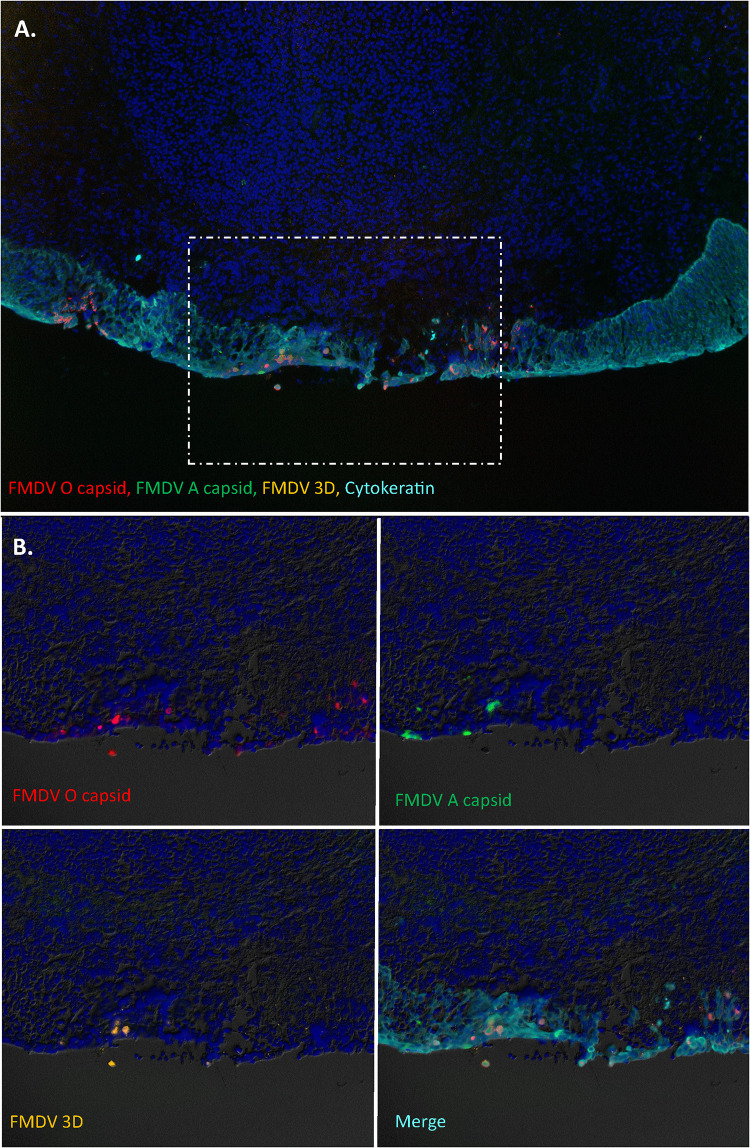
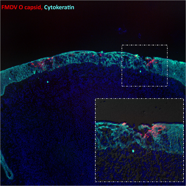
Superinfection of cattle persistently infected with foot-and-mouth disease virus (FMDV), with a heterologous FMDV strain has been shown to generate novel recombinant viruses. In this study, we investigated the pathogenesis events within specific tissues associated with FMDV coinfections in cattle subjected to either simultaneous or serial exposure to two distinct strains of FMDV. Both strains of FMDV (one each of serotypes O and A) were similarly localized to the nasopharyngeal mucosa during the early stages of infection. However, while no recombinant FMDV genomes were recovered from simultaneously coinfected cattle, interserotypic recombinants were isolated from nasopharyngeal tissue samples obtained at 48 h after heterologous superinfection of a persistently infected FMDV carrier. Additionally, analysis of FMDV genomes obtained from replicate nasopharyngeal tissue samples demonstrated that adjacent segments of the mucosa were sometimes infected by distinct viruses, demonstrating a multifocal and heterogeneous distribution of FMDV infection during primary and persistent phases of infection. This work indicates that superinfection of FMDV carriers may be an important source of emergent recombinant strains of FMDV in areas where multiple strains are co-circulating. IMPORTANCE Foot-and-mouth disease (FMD) is a socioeconomically impactful livestock disease with a complex epidemiology and ecology. Although recombinant viruses have been identified in field samples, the mechanisms of emergence of those viruses have never been elucidated. This current study demonstrates how serial infection of cattle with two distinct serotypes of FMD virus (FMDV) leads to rapid generation of recombinant viruses in the upper respiratory tracts of infected animals. This finding is particularly relevant in relation to the management of persistently infected FMDV carrier cattle that can maintain subclinical FMDV infection for months to years after an initial infection. Such carrier animals may function as mixing vessels that facilitate the emergence of novel recombinant FMDV strains in areas where multiple virus strains are in circulation.

Keywords: FMD; FMDV; cattle; foot-and-mouth disease; foot-and-mouth disease virus; infectious disease; pathogenesis; recombination.


Conflict of interest statement

The authors declare no conflict of interest.

Figures

FIG 1
FIG 1
Study design. The study comprised three groups of four cattle each. Group 1 were simultaneously infected with foot-and-mouth disease virus (FMDV) O1 Manisa and A24 Cruzeiro on day 0, Group 2 were infected with FMDV A24 Cruzeiro on day 0 and superinfected with FMDV O1 Manisa on day 21, and Group 3 were infected with FMDV A24 Cruzeiro on day 0 and superinfected with FMDV O1 Manisa on day 35. Two animals from each group were euthanized for postmortem tissue harvest at 24 and 48 h after the final (or, for group 1, simultaneous) virus exposure.
FIG 2
FIG 2
FMDV infection dynamics in simultaneously coinfected cattle. Strain-specific detection of FMDV RNA in nasal swabs (solid lines, triangular markers) and sera (hatched lines, circular markers) from cattle simultaneously infected with FMDV O1 Manisa (red) and FMDV A24 Cruzeiro (blue). Animals no. 19-01 and 19-02 (top row) were euthanized for tissue harvest at 1 day postinfection (dpi) and animals no. 19-03 and 19-04 (bottom row) were euthanized at 2 dpi. No FMD lesions were observed in any of the animals.
FIG 3
FIG 3
FMDV distribution in bovine tissues following simultaneous coinfection. Strain-specific FMDV detection in tissue samples obtained at 24 and 48 h post-intranasopharyngeal deposition of a mixed inoculum containing equal quantities of FMDV O1 Manisa (red) and FMDV A24 Cruzeiro (blue). Numbers in table represent log10 genome copy numbers (GCN)/mg of FMDV RNA in tissue or log10 GCN/μL serum, for the specific virus. Color gradient indicates increasing FMDV RNA quantities in samples that were positive by both strain-specific real-time reverse transcriptase PCR (qRT-PCR) and virus isolation (VI), with virus specificity in VI supernatants determined by strain-specific qRT-PCR. Numbers in uncolored cells indicate quantities of FMDV RNA detected in samples that were negative by virus isolation for that specific virus; plus sign (+) indicates that virus isolation was positive but FMDV RNA content was below the limit of detection.
FIG 4
FIG 4
FMDV infection dynamics in cattle that were infected with FMDV A24 Cruzeiro on day 0 and superinfected with FMDV O1 Manisa on day 21. Strain-specific detection of FMDV RNA in nasal swabs (solid lines, triangular markers), sera (hatched lines, circular markers), and oropharyngeal fluid (OPF; purple line, diamond markers) from cattle sequentially infected with FMDV A24 Cruzeiro (blue and purple) and FMDV O1 Manisa (red). Shaded yellow area represents the cumulative lesion score observed through the first 10 days of infection. Animals no. 19-09 and 19-10 (top row) were euthanized for tissue harvest at 1 day post-superinfection, and animals no. 19-11 and 19-12 (bottom row) were euthanized at 2 days post-superinfection. Asterisk (*) indicates that OPF samples were only collected on days 14 and 17.
FIG 5
FIG 5
FMDV infection dynamics in cattle that were infected with FMDV A24 Cruzeiro on day 0 and superinfected with FMDV O1 Manisa on day 35. Strain-specific detection of FMDV RNA in nasal swabs (solid lines, triangular markers), sera (hatched lines, circular markers), and OPF (solid purple line, diamond markers) from cattle sequentially infected with FMDV A24 Cruzeiro (blue and purple) and FMDV O1 Manisa (red). Shaded yellow area represents the cumulative lesion score observed through the first 10 days of infection. Animals no. 19-33 and 19-34 (top row) were euthanized for tissue harvest at 1 day post-superinfection, and animals no. 19-35 and 19-36 (bottom row) were euthanized at 2 days post superinfection. Asterisk (*) indicates that OPF samples were collected twice weekly from days 14 to 31.
FIG 6
FIG 6
FMDV distribution in bovine tissues following FMDV O1 Manisa (red) superinfection, 21 days after initial infection with FMDV A24 Cruzeiro (blue). Strain-specific FMDV detection in tissue samples obtained 24 and 48 h post-FMDV O1 Manisa superinfection. Numbers in table represent log10 GCN/mg of FMDV RNA in tissue or log10 GCN/μL serum for the specific virus. Color gradient indicates increasing FMDV RNA quantities in samples that were positive by both strain-specific qRT-PCR and VI, with virus specificity in VI supernatants determined by strain-specific qRT-PCR. Numbers in uncolored cells indicate quantities of FMDV RNA detected in samples that were negative by virus isolation for that specific virus; plus sign (+) indicates that virus isolation was positive but FMDV RNA content was below the limit of detection.
FIG 7
FIG 7
FMDV distribution in bovine tissues following FMDV O1 Manisa (red) superinfection, 35 days after initial infection with FMDV A24 Cruzeiro (blue). Strain-specific FMDV detection in tissue samples obtained 24 and 48 h post-FMDV O1 Manisa superinfection. Numbers in the table represent log10 GCN/mg of FMDV RNA in tissue or log10 GCN/μL serum for the specific virus. Color gradient indicates increasing FMDV RNA quantities in samples that were positive by both strain-specific qRT-PCR and VI, with virus specificity in VI supernatants determined by strain-specific qRT-PCR. Numbers in uncolored cells indicate quantities of FMDV RNA detected in samples that were negative by virus isolation for that specific virus; plus sign (+) indicates that virus isolation was positive but FMDV RNA content was below the limit of detection.
FIG 8
FIG 8
FMDV coinfection in the bovine nasopharyngeal mucosa. (A) FMDV infection in the dorsal nasopharyngeal mucosa at 24 h post-intranasopharyngeal deposition of a mixed inoculum containing equal quantities of FMDV O1 Manisa and FMDV A24 Cruzeiro. (B) FMDV VP1 (O1 Manisa = red, A24 Cruzeiro = green) is localized to cytokeratin+ epithelial cells (teal) within a surface erosion in a segment of follicle-associated epithelium. FMDV 3D protein (orange) was detected in a cluster of a few cells within the same region. There is minimal to no colocalization of FMDV O1M and A24. Panel A = ×10 magnification. Panel B = ×40 magnification with differential interference contrast of region of interest showing separate and merged channels.
FIG 9
FIG 9
FMDV O1 Manisa superinfection in bovine nasopharynx persistently infected with FMDV A24 Cruzeiro. (A) Early-phase FMDV O1 Manisa infection in the dorsal nasopharyngeal mucosa at 48 h post-FMDV O1 Manisa superinfection of an animal that was persistently infected with FMDV A24 Cruzeiro. (B) FMDV O1 Manisa VP1 (red) is associated with multiple surface erosions within segments of follicle-associated epithelium (cytokeratin = teal). FMDV A24 VP1 (green) is localized to a single cluster of cytokeratin+ epithelial cells directly adjacent to an epithelial erosion. FMDV 3D protein (orange) was detected in a few cells within the same region. Panel A = ×10 magnification. Panel B = ×20 magnification with differential interference contrast of region of interest showing separate and merged channels.
FIG 10
FIG 10
Acute FMDV O1 Manisa infection in the bovine nasopharyngeal mucosa. Multiple foci of FMDV O1 Manisa (red) infection associated with erosions within the mucosal surface of the dorsal soft palate at 48 h postinfection. The VP1 antigen is exclusively detected within cytokeratin+ (teal) cells within segments of follicle-associated epithelium. Image = ×10 magnification. Inset: ×40 magnification with differential interference contrast of region of interest.
FIG 11
FIG 11
Characterization of FMDV infection by next-generation sequencing (NGS) in distinct compartments of the bovine nasopharynx. Viruses isolated from four distinct compartments of the nasopharynx: rostral and caudal segments of the dorsal surface of the soft palate as well as rostral and caudal segments of the dorsal nasopharynx were analyzed by NGS. All samples obtained from simultaneously infected animals (Group 1) contained both viruses (purple cells). In animals which had been superinfected with FMDV O1 Manisa 21 or 35 days post-infection with FMDV A24 (Groups 2 and 3), FMDV O1 Manisa (red cells) had a wider distribution compared to FMDV A24. Only one tissue replicate contained FMDV A24 without concurrent detection of FMDV O1M (blue cell). Split cells of different color designations indicate that different viruses, or combinations of viruses, were detected in replicate samples within the same anatomic compartment. Asterisk(*) indicates that samples from animal no. 19-36 contained an interserotypic recombinant virus which was confirmed through plaque purification and analysis of plaques by NGS.
FIG 12
FIG 12
Schematic of the interserotypic recombinant FMDV genome isolated from the dorsal soft palate of animal no. 19-36, obtained 48 h post-FMDV O1M superinfection. The virus identified in four sequenced plaques was comprised of an FMDV O1M-derived genome from the 5′ UTR through the center of the 3D coding region and an FMDV A24-derived coding region from the mid-3D through the 3′ UTR. In each alignment, horizontal rows colored light grey indicate identity with the reference sequence while black banding indicates dissimilarity.

Similar articles

Cited by

References

    1. Grubman MJ, Baxt B. 2004. Foot-and-mouth disease. Clin Microbiol Rev 17:465–493. doi:10.1128/CMR.17.2.465-493.2004. - DOI - PMC - PubMed
    1. Office International des Epizooties (OIE). 2022. Application for official recognition by the OI of free status for foot-and-mouth disease virus. In Terrestrial Animal Health Code. Available from https://www.woah.org/fileadmin/Home/eng/Health_standards/tahc/current/ch.... WOAH, Paris, France.
    1. Shanafelt DW, Perrings CA. 2017. Foot and mouth disease: the risks of the international trade in live animals. Rev Sci Tech 36:839–865. doi:10.20506/rst.36.3.2719. - DOI - PubMed
    1. Knight-Jones TJ, Rushton J. 2013. The economic impacts of foot and mouth disease: what are they, how big are they and where do they occur? Prev Vet Med 112:161–173. doi:10.1016/j.prevetmed.2013.07.013. - DOI - PMC - PubMed
    1. Knight-Jones TJ, McLaws M, Rushton J. 2017. Foot-and-mouth disease impact on smallholders: what do we know, what don’t we know and how can we find out more? Transbound Emerg Dis 64:1079–1094. doi:10.1111/tbed.12507. - DOI - PMC - PubMed

LinkOut - more resources