. 2023 Apr 3;64(4):28.
doi: 10.1167/iovs.64.4.28.

Effects of Plasma-Derived Exosomal miRNA-19b-3p on Treg/T Helper 17 Cell Imbalance in Behçet's Uveitis

Affiliations

Effects of Plasma-Derived Exosomal miRNA-19b-3p on Treg/T Helper 17 Cell Imbalance in Behçet's Uveitis

Qingyan Jiang et al. Invest Ophthalmol Vis Sci. .

Abstract

Purpose: To explore the potential role of plasma-derived exosomal microRNAs (miRNAs) in the development of regulatory T cell (Treg)/T helper 17 (Th17) cell imbalances in Behçet's uveitis (BU).

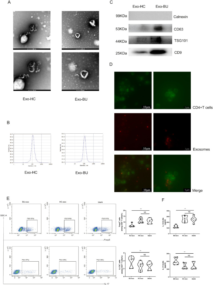
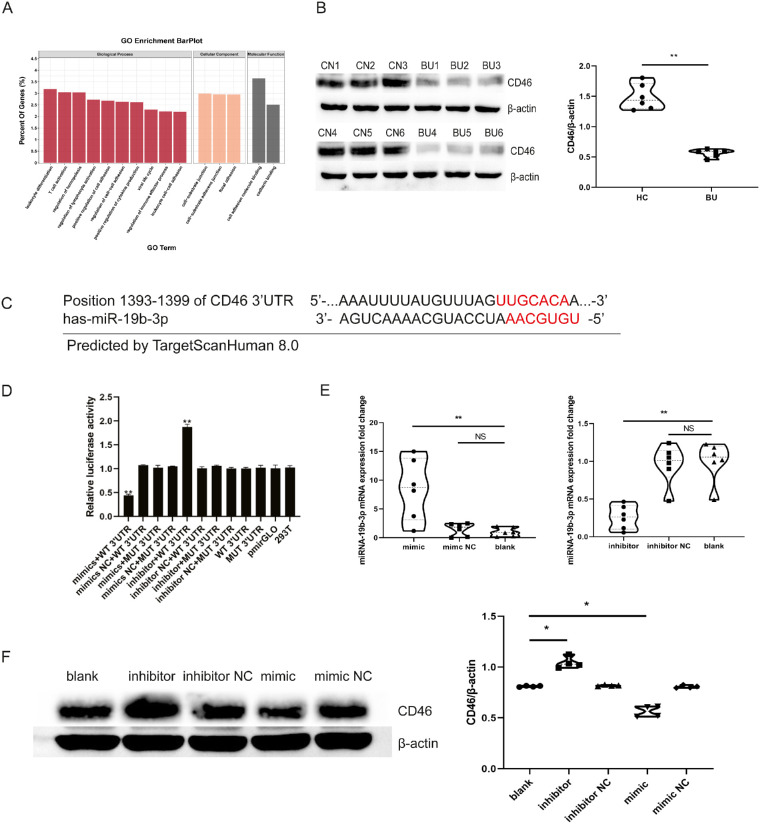
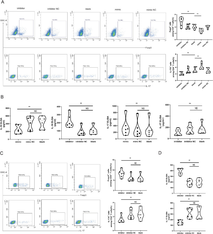
Methods: The exosome treatment was conducted to evaluate the effects of plasma exosomes from patients with active BU and healthy controls on the Treg/Th17 cell balance. miRNA sequencing analysis of plasma exosomes was conducted to identify differentially expressed miRNAs between patients with active BU and healthy controls. miRTarBase analysis and dual-luciferase reporter assays were conducted to identify the target genes of miR-19b-3p. CD4+T cells were transfected with miR-19b-3p mimic or inhibitor to evaluate its regulation of the Treg/Th17 cell balance. The Treg/Th17 cell balance in CD4+T cells was evaluated by flow cytometry and enzyme-linked immunosorbent assay.

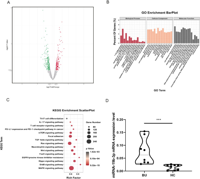
Results: Exosomes from patients with active BU promoted Th17 cell differentiation and inhibited Treg cell differentiation. MiRNA sequencing analysis revealed 177 upregulated and 274 downregulated miRNAs in plasma exosomes of patients with active BU. Among them, miR-19b-3p was significantly elevated, and its target genes were identified as being involved in T-cell differentiation. miR-19b-3p overexpression downregulated CD46 expression and the Treg/Th17 cell ratio in CD4+T cells from healthy controls, whereas miR-19b-3p inhibition reversed these regulatory effects and restored the Treg/Th17 cell balance of CD4+T cells from patients with active BU.

Conclusions: Plasma-derived exosomes from patients with active BU showed a markedly differential miRNA expression in comparison to healthy controls. Highly expressed miRNA-19b-3p could induce a Treg/Th17 cell imbalance, probably by downregulating CD46 expression.


Conflict of interest statement

Disclosure: Q. Jiang, None; Q. Wang, None; S. Tan, None; J. Cai, None; X. Ye, None; G. Su, None; P. Yang, None

Figures

Figure 1.
Figure 1.
Exosomes from patients with active BU induced a Treg/Th17 cell imbalance. (A) Exosomes were isolated from plasma of healthy controls and patients with active BU, and their morphology and size were validated by TEM. Scale bars: 200 nm and 100 nm. (B) NTA was used to evaluate the size and concentration of plasma exosomes. (C) WB analysis was used to detect positive exosome markers (CD9, CD63, and TSG101) and a negative exosome marker (calnexin). (D) Representative fluorescence microscopy images showing the internalization of PKH-26-labeled exosomes (red) by PKH-67-labeled CD4+T cells (green). Scale bars: 10 µm and 1 µm. (E) The proportion of Treg and Th17 in CD4+T cells was detected by FCM following 24 hours of treatment with plasma exosomes from patients with active BU, plasma exosomes from healthy controls, or PBS (n = 6 for each treatment). (F) The protein levels of IL-10 and IL-17 were measured by ELISA following 24 hours of treatment with plasma exosomes from patients with active BU, plasma exosomes from healthy controls, or PBS (n = 6 for each treatment). All experiments were repeated three times. The paired ANOVA test was used for statistical analysis (*P < 0.05, **P < 0.01); NS, no significance.
Figure 2.
Figure 2.
The exosomal miRNA profiles of patients with active BU differed from those of healthy controls. (A) Volcano plot comparing expressed exosomal miRNAs in plasma from patients with active BU and from healthy controls. A volcano plot was created using a log2 fold change and –log10 P values of all of the detected miRNAs. (B) GO pathway enrichment analysis of the target genes associated with the differentially expressed miRNAs was performed, and 45 pathways related to biological processes (BPs), cellular components (CCs), and molecular functions (MFs) were identified. (C) KEGG analysis of differential miRNA target genes identified 15 immune-related pathways. (D) Exosomal miR-19b-3p expression in plasma of patients with active BU and healthy controls as detected by RT-qPCR. The patients with active BU did not use immunosuppressive drugs for at least 1 week or used less than 20 mg/d prednisone less than 1 week before blood sampling (n = 8 for each treatment). Experiments were repeated three times. Data were analyzed using an unpaired t-test (***P < 0.001).
Figure 3.
Figure 3.
CD46 was the target gene of exosomal miR-19b-3p. (A) GO pathway enrichment analysis of CD46 identified 15 pathways related to BPs, CCs, and MFs. (B) Comparison of CD46 protein expression in CD4+T cells from patients with active BU and healthy controls (n = 6 for each treatment). (C) miR-19b-3p binding sites in the CD46 3′ UTR. The binding sequence of miR-19b-3p is located at nucleotides 1393 to 1399 from the 3′ UTR, as predicted by TargetScanHuman 8.0. (D) The relative luciferase activity of the 3′ UTR-CD46-WT or 3′ UTR-CD46-MUT vectors in 293T cells co-transfected with miR-19b-3p mimic or NC mimic or with miR-19b-3p inhibitor or NC inhibitor. (E) The mRNA levels of miR-19b-3p in CD4+T cells transfected with miR-19b-3p mimic or NC mimic or with miR-19b-3p inhibitor or NC inhibitor measured by RT-qPCR (n = 6 for each treatment). (F) WB analysis of CD46 expression in CD4+T cells transfected with miR-19b-3p mimic or NC mimic or with miR-19b-3p inhibitor or NC inhibitor (n = 4 for each treatment). All experiments were repeated three times. P values were calculated using the paired t-test to compare two groups. For multiple comparisons, P values were calculated using the paired ANOVA test (*P < 0.05, **P < 0.01); NS, no significance.
Figure 4.
Figure 4.
miR-19b-3p transfection in CD4+T cells increased Th17 cell frequency and IL-17 levels while decreasing Treg cell frequency and IL-10 levels. (A) The proportions of Treg and Th17 in CD4+T cells transfected with miR-19b-3p mimic or NC mimic or with miR-19b-3p inhibitor or NC inhibitor were detected by FCM (n = 6 for each treatment). (B) The protein levels of IL-10 and IL-17 in CD4+T cells transfected with miR-19b-3p mimic or NC mimic or with miR-19b-3p inhibitor or NC inhibitor were measured by ELISA (n = 6 for each treatment). (C) The proportions of Treg and Th17 in CD4+T cells from patients with active BU transfected with miR-19b-3p inhibitor or NC inhibitor were detected by FCM (n = 6 for each treatment). (D) The protein levels of IL-10 and IL-17 in CD4+T cells from patients with active BU transfected with miR-19b-3p inhibitor or NC inhibitor were measured by ELISA (n = 6 for each treatment). All experiments were repeated three times. Data are presented as the mean ± SEM of three independent experiments. For multiple comparisons, the paired ANOVA test was applied (*P < 0.05, **P < 0.01); NS, no significance.

References

    1. Deuter CM, Kötter I, Wallace GR, Murray PI, Stübiger N, Zierhut M.. Behçet's disease: Ocular effects and treatment. Prog Retin Eye Res. 2008; 27(1): 111–136. - PubMed
    1. Yang P, Ohno S, Zierhut M. Editorial: New insights into uveitis: Immunity, genes, and microbes. Front Immunol. 2021; 12: 765377. - PMC - PubMed
    1. Triolo G, Accardo-Palumbo A, Dieli F, et al. .. Humoral and cell mediated immune response to cow's milk proteins in Behçet's disease. Ann Rheum Dis. 2002; 61(5): 459–462. - PMC - PubMed
    1. Mendes D, Correia M, Barbedo M, et al. .. Behçet's disease – a contemporary review. J Autoimmun. 2009; 32: 178–188. - PubMed
    1. Cunningham ET Jr, Tugal-Tutkun I, Khairallah M, Okada AA, Bodaghi B, Zierhut M. Behçet uveitis. Ocul Immunol Inflamm. 2017; 25(1): 2–6. - PubMed

Publication types