Meta-Analysis
. 2023 May;228(3-4):997-1017.
doi: 10.1007/s00429-023-02641-y. Epub 2023 Apr 24.

Accurate localization and coactivation profiles of the frontal eye field and inferior frontal junction: an ALE and MACM fMRI meta-analysis

Affiliations
Meta-Analysis

Accurate localization and coactivation profiles of the frontal eye field and inferior frontal junction: an ALE and MACM fMRI meta-analysis

Marco Bedini et al. Brain Struct Funct. 2023 May.

Abstract

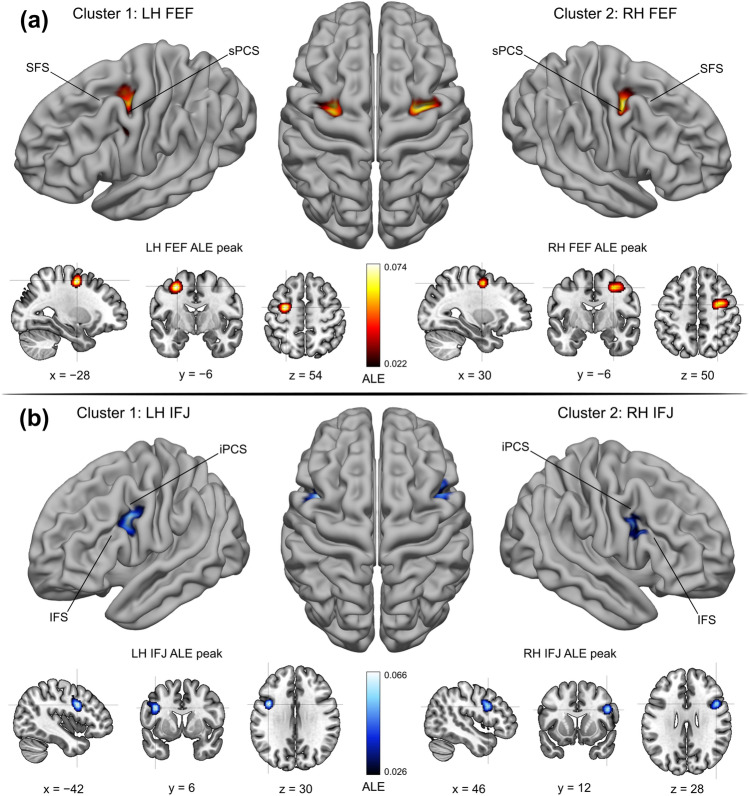
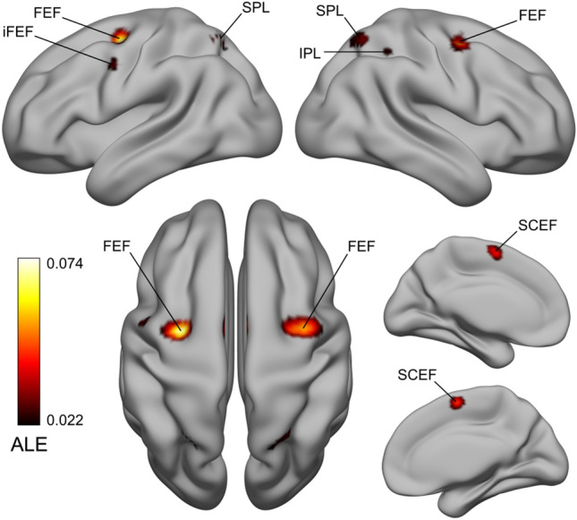
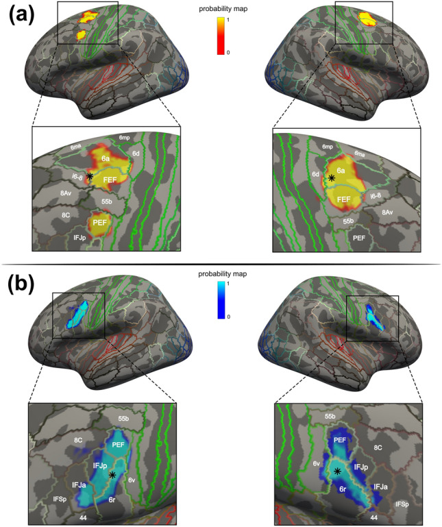
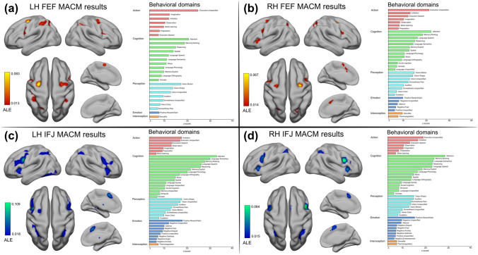
The frontal eye field (FEF) and the inferior frontal junction (IFJ) are prefrontal structures involved in mediating multiple aspects of goal-driven behavior. Despite being recognized as prominent nodes of the networks underlying spatial attention and oculomotor control, and working memory and cognitive control, respectively, the limited quantitative evidence on their precise localization has considerably impeded the detailed understanding of their structure and connectivity. In this study, we performed an activation likelihood estimation (ALE) fMRI meta-analysis by selecting studies that employed standard paradigms to accurately infer the localization of these regions in stereotaxic space. For the FEF, we found the highest spatial convergence of activations for prosaccade and antisaccade paradigms at the junction of the precentral sulcus and superior frontal sulcus. For the IFJ, we found consistent activations across oddball/attention, working memory, task-switching and Stroop paradigms at the junction of the inferior precentral sulcus and inferior frontal sulcus. We related these clusters to previous meta-analyses, sulcal/gyral neuroanatomy, and a comprehensive brain parcellation, highlighting important differences compared to their results and taxonomy. Finally, we leveraged the ALE peak coordinates as seeds to perform a meta-analytic connectivity modeling (MACM) analysis, which revealed systematic coactivation patterns spanning the frontal, parietal, and temporal cortices. We decoded the behavioral domains associated with these coactivations, suggesting that these may allow FEF and IFJ to support their specialized roles in flexible behavior. Our study provides the meta-analytic groundwork for investigating the relationship between functional specialization and connectivity of two crucial control structures of the prefrontal cortex.

Keywords: Activation likelihood estimation; Cognitive control; Meta-analytic connectivity modeling; Prefrontal cortex; Saccades; Working memory.


Conflict of interest statement

The authors have no relevant financial or non-financial interests to disclose.

Figures

Fig. 1
Fig. 1
FEF and IFJ localizer samples—ALE main clusters. A ALE results from the FEF localizer sample. Two main clusters were found in the posterior dorsolateral PFC, which corresponds to the description of the anatomical location of the FEF (Paus ; Vernet et al. 2014). The FEF peaks were localized at the junction of the sPCS and the SFS, in the anterior (in the LH) and posterior (in the RH) banks of the sPCS. B ALE results from the IFJ localizer sample. Two main clusters were found in the posterior ventrolateral PFC, and their respective peaks were localized along the posterior bank of the iPCS, near its ventral junction with the IFS. The location of these peaks and the corresponding MNI152 coordinates match the description of the IFJ (Derrfuss et al. ; Muhle-Karbe et al. 2016)
Fig. 2
Fig. 2
FEF localizer sample ALE results—FEF lateral peak and other significant clusters. In the FEF localizer analysis, we also found a lateral peak in the LH, which was localized near the bank of the iPCS, dorsal to its junction with the IFS, corresponding to the iFEF (Derrfuss et al. ; Kastner et al. 2007). It is unclear whether this region should be considered part of the FEF proper (Glasser et al. ; Mackey et al. 2017). We found three other significant clusters localized in the supplementary and cingulate eye field (SCEF) and the dorsal cingulate motor cortex, and the precuneus/superior parietal lobule (SPL) and the right anterior intraparietal area. These areas form some of the core control structures of the oculomotor network (Coiner et al. 2019)
Fig. 3
Fig. 3
IFJ localizer sample ALE results—Other significant clusters. In addition to the bilateral IFJ clusters, we found significant activations in the dACC/SCEF, the left FEF, in two clusters in the insular cortex and claustrum (not visible in the LH), and finally, in the SPL/IPL. Given that these areas were activated across different paradigms, we suggest that they could be associated with the “encoding and updating of task-relevant representations” as first hypothesized by Derrfuss et al. (; see also Rodríguez-Nieto et al. 2022). These areas mostly belong to the frontoparietal network (Yeo et al. 2011)
Fig. 4
Fig. 4
Projection on FSaverage of the FEF and IFJ main clusters and comparison with the MMP1. A Vertices corresponding to the FEF clusters. Both clusters covered the middle and rostral parts of the FEF label as defined according to the MMP1 atlas, but they also covered large parts of area 6a. In the LH, vertices were also localized in the iFEF, which matches almost exactly the boundaries of area PEF from the atlas. The LH FEF ALE peak was localized within area i6-8, just anterior to the FEF, and the RH FEF ALE peak was localized within area 6a, dorsal to FEF. Despite this difference, both peaks were localized near the junction of the sPCS and the SFS, in the anterior bank and the posterior banks of the sPCS, respectively. B Vertices corresponding to the IFJ clusters. They showed a similar elongated shape that approximately followed the posterior iPCS and encroached onto the IFS, and they spanned multiple MMP1 areas. Importantly, we found that in both hemispheres the IFJ ALE peaks were localized near the junction of the iPCS and the SFS within area 6r, posteriorly to the IFJp
Fig. 5
Fig. 5
Meta-analytic connectivity modeling results. A and B depict the coactivation profiles of the FEF, and C and D depict the coactivation profiles of the IFJ. On the right side of each panel: decoding results of the significant associations with behavioral domains (p < 0.05, Bonferroni corrected)

References

    1. Abdollahi RO, Kolster H, Glasser MF, et al. Correspondences between retinotopic areas and myelin maps in human visual cortex. Neuroimage. 2014;99:509–524. doi: 10.1016/j.neuroimage.2014.06.042. - DOI - PMC - PubMed
    1. Amiez C, Petrides M. Anatomical organization of the eye fields in the human and non-human primate frontal cortex. Prog Neurobiol. 2009;89:220–230. doi: 10.1016/j.pneurobio.2009.07.010. - DOI - PubMed
    1. Amiez C, Petrides M. Functional rostro-caudal gradient in the human posterior lateral frontal cortex. Brain Struct Funct. 2018;223:1487–1499. doi: 10.1007/s00429-017-1567-z. - DOI - PubMed
    1. Amiez C, Kostopoulos P, Champod AS, Petrides M. Local morphology predicts functional organization of the dorsal premotor region in the human brain. J Neurosci. 2006;26:2724–2731. doi: 10.1523/JNEUROSCI.4739-05.2006. - DOI - PMC - PubMed
    1. Amunts K, Von Cramon DY. Special issue: position paper the anatomical segregation of the frontal cortex: what does it mean for function. Brain. 2006;3:525–528. doi: 10.1016/S0010-9452(08)70392-7. - DOI - PubMed

Publication types